44 light microscope with labels
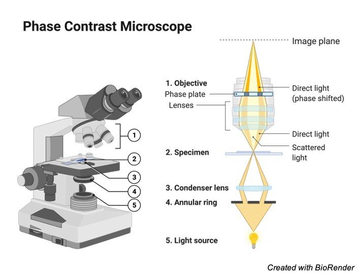
› microscopy › intSmart Microscope for Lab Routine and Research - ZEISS Acquiring fluorescent images has never been so easy. Combine Axioscope 5 with the LED light source Colibri 3 and the sensitive, standalone microscope camera Axiocam 202 mono to have the perfect setup for easy multichannel fluorescence documentation. Switch effortlessly between the channels for UV, blue, green and red excitation. An Introduction to the Light Microscope, Light Microscopy Techniques ... Figure 1: Basic compound microscope: Light from a source is focused onto the sample (object) using a mirror and condenser lens. Light from the sample is collected by an objective, forming an intermediate image which is imaged again by the eyepiece and relayed to the eye, which sees a magnified image of the sample.
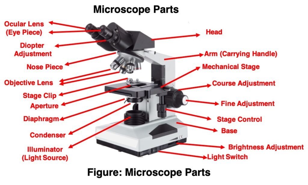
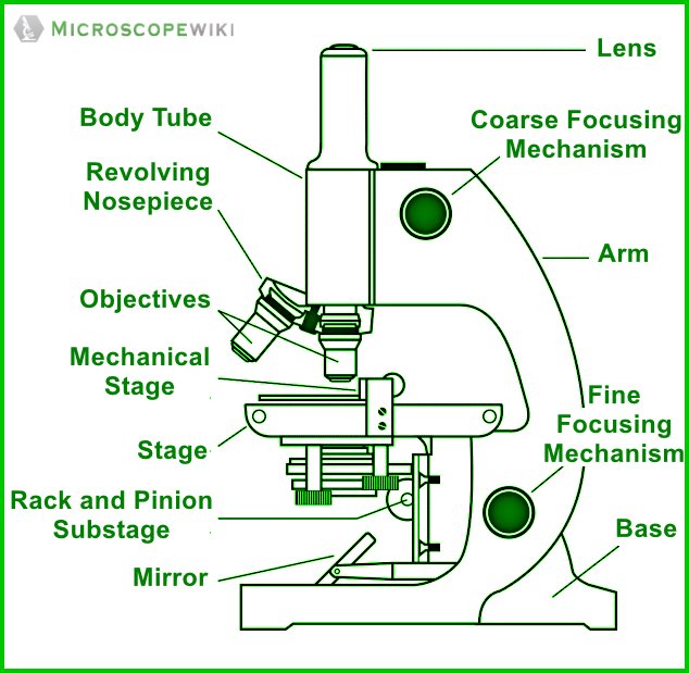
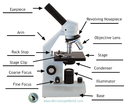
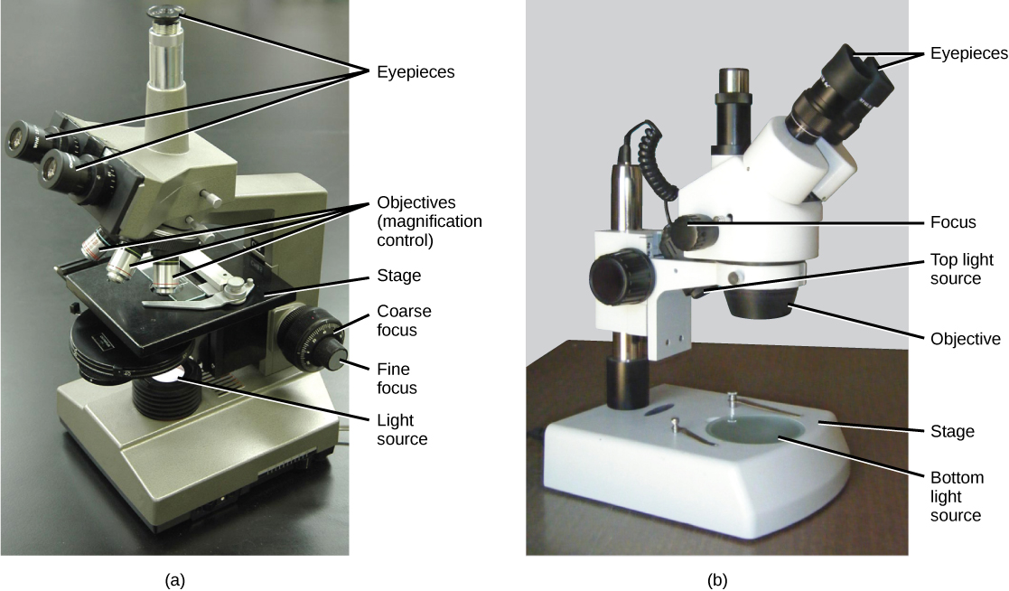
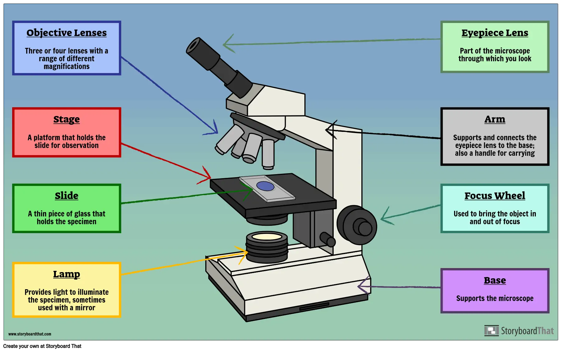
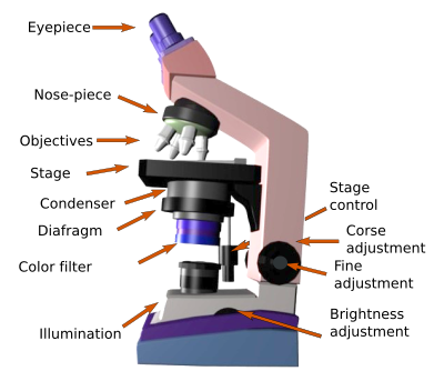
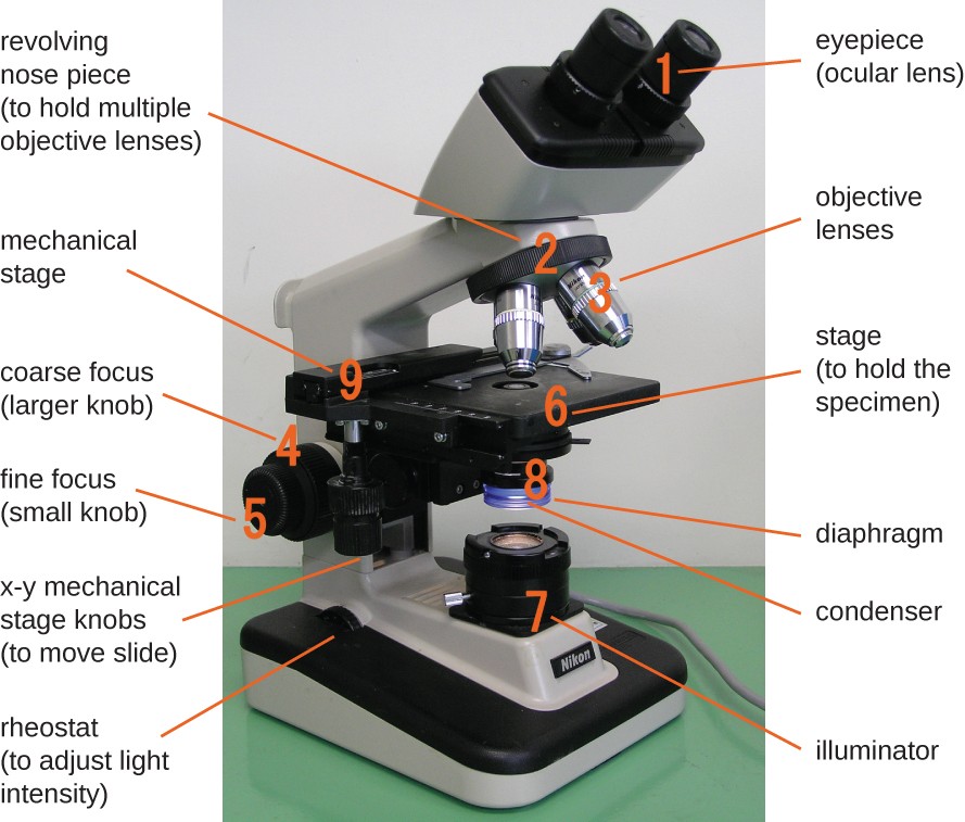
Compound Microscope Parts, Functions, and Labeled Diagram Abbe Condenser: This lens condenses the light from the base illumination and focuses it onto the stage. This piece of the compound microscope sits below the stage & typically acts as a structural support that connects the stage to arm or frame of the microscope. Coarse and fine adjustment controls: Adjusts the focus of the microscope.
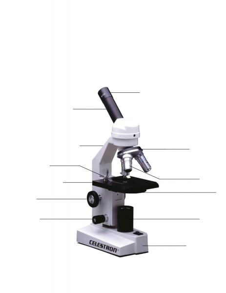
Light microscope with labels
› cemf › whatisemWhat is Electron Microscopy? - UMASS Medical School Conventional scanning electron microscopy depends on the emission of secondary electrons from the surface of a specimen. Because of its great depth of focus, a scanning electron microscope is the EM analog of a stereo light microscope. It provides detailed images of the surfaces of cells and whole organisms that are not possible by TEM. Required practical - using a light microscope - BBC Bitesize Record the microscope images using labelled diagrams or produce digital images. When first examining cells or tissues with low power, draw an image at this stage, even if going on to examine the... A Study of the Microscope and its Functions With a Labeled Diagram These labeled microscope diagrams and the functions of its various parts, attempt to simplify the microscope for you. However, as the saying goes, 'practice makes perfect', here is a blank compound microscope diagram and blank electron microscope diagram to label.
Light microscope with labels. Polarizing Microscope Image Gallery - Leica Microsystems Polarized light microscopy (also known as polarizing microscopy) is an important method used in different fields, including research and quality assurance. It goes beyond just producing images at high magnification and resolution, something typically done with microscopes using ordinary optics. By examining the form, structure, color, birefringence, and further optical properties, additional ... Labeling Microscope Worksheet | Teaching Resources A straightforward worksheet in which students are required to identify the parts of a basic microscope. Tes classic free licence. Report this resource to let us know if it violates our terms and conditions. Our customer service team will review your report and will be in touch. Last updated. 21 November 2014. rsscience.com › stereo-microscopeParts of Stereo Microscope (Dissecting microscope) - Rs' Science Stereo microscopes (also called Dissecting microscope) are branched out from other light microscopes for the application of viewing "3D" objects. These include substantial specimens, such as insects, feathers, leaves, rocks, sand grains, gems, coins, and stamps, etc. Functionally, a stereo microscope is like a powerful magnifying glass. Light Microscopy Virtual Lab - Labster About This Simulation. This simulation, along with "Fluorescence Microscopy," has been adapted from the original, larger "Microscopy" simulation. Assemble the light microscope and discover how the key components help to magnify an image up to 1,000 times. In this simulation, you will learn how to use a light microscope to analyze an ...
Simple Microscope - Parts, Functions, Diagram and Labelling Parts of the optical parts are as follows: Mirror - A simple microscope has a plano-convex mirror and its primary function is to focus the surrounding light on the object being examined. Lens - The biconvex lens is placed above the stage and its function is to magnify the size of the object being examined. Light microscopes - Cell structure - Edexcel - BBC Bitesize Microscopes are used to produce magnified images. There are two main types of microscope: light microscopes are used to study living cells and for regular use when relatively low magnification and... Parts of a microscope with functions and labeled diagram The higher the magnification of the condenser, the more the image clarity. More sophisticated microscopes come with an Abbe condenser that has a high magnification of about 1000X. Diaphragm - it's also known as the iris. Its found under the stage of the microscope and its primary role is to control the amount of light that reaches the specimen. Labeling the Parts of the Microscope Labeling the Parts of the Microscope This activity has been designed for use in homes and schools. Each microscope layout (both blank and the version with answers) are available as PDF downloads. You can view a more in-depth review of each part of the microscope here. Download the Label the Parts of the Microscope PDF printable version here.
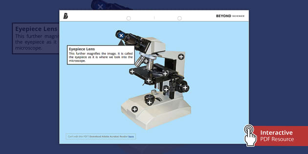
Compound Microscope Parts - Labeled Diagram and their Functions The eyepiece (or ocular lens) is the lens part at the top of a microscope that the viewer looks through. The standard eyepiece has a magnification of 10x. You may exchange with an optional eyepiece ranging from 5x - 30x. [In this figure] The structure inside an eyepiece. The current design of the eyepiece is no longer a single convex lens. Fluorescence Microscopy - Explanation and Labelled Images A fluorescence microscope is used to study organic and inorganic samples. Fluorescence microscopy uses fluorescence and phosphorescence to examine the structural organization, spatial distribution of samples. It is particularly used to study samples that are complex and cannot be examined under conventional transmitted-light microscope. en.wikipedia.org › wiki › Two-photon_excitationTwo-photon excitation microscopy - Wikipedia In scattering tissue, on the other hand, the superior optical sectioning and light detection capabilities of the two-photon microscope result in better performance. Applications Main. Two-photon microscopy has been involved with numerous fields including: physiology, neurobiology, embryology and tissue engineering. Researchers demonstrate label-free super-resolution microscopy A newly developed sub-diffraction-limit microscopy approach doesn't require fluorescent labels. The video shows the process of the data evaluation algorithm, retrieving the positions and sizes of...
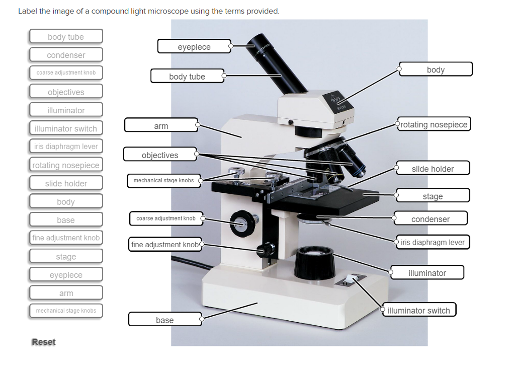
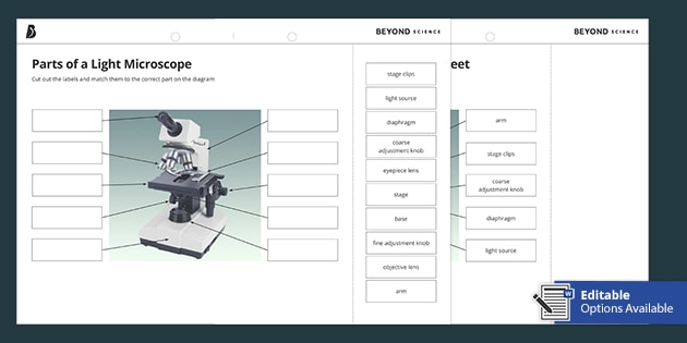
› 6-label-the-microscopeLabel the microscope — Science Learning Hub All microscopes share features in common. In this interactive, you can label the different parts of a microscope. Use this with the Microscope parts activity to help students identify and label the main parts of a microscope and then describe their functions. Drag and drop the text labels onto the microscope diagram. If you want to redo an answer, click on the box and the answer will go back to the top so you can move it to another box.
Microscope, Microscope Parts, Labeled Diagram, and Functions Majority of high quality microscopes used in laboratory include an Abbe condenser with an iris diaphragm. When iris diaphragm is combined with Abbe condenser, it control both the quantity of light applied as well as focus on the specimen. Aperture: It is the hole in the stage through which the base (transmitted) light reaches the stage.
Cell Under Labeled Microscope Leaf The microscope is an normal level microscope,it is not good at living cells analysis 2mm, Built-in WF 16mm Eyepiece - Capture and Record The Beauty in The Micro World Microscopes are an essential tool to use when studying cells 7-2mm broad with serrations or small spines along the leaf margins Procedure 1 Place a hydrilla sprig in a test tube ...
› microscopy › intLightsheet 7 - Light Sheet Microscope to Image Live or ... What’s more, use this light sheet microscope to image very large optically cleared specimens in toto, and with subcellular resolution. Enhance your Lightsheet 7 with dedicated optics, sample chambers and sample holders to accurately adjust to the refractive index of your chosen clearing method, and then image your large samples, even whole ...
Light Microscopy - an overview | ScienceDirect Topics Optical microscopy enables minimally invasive imaging of cellular structures and processes with high specificity via fluorescent labeling, but its spatial resolution is fundamentally limited to approximately half the wavelength of light. Structured illumination microscopy (SIM) can improve this limit by a factor of two while keeping all ...
Compound Light Microscopes | Products | Leica Microsystems Leica Microsystems provides microscope parts and accessories to perfectly tailor your imaging system for your needs and budget. Darkfield Microscopes A darkfield microscope offers a way to view the structures of many types of biological specimens in greater contrast without the need of stains. Products Leica Science Lab Key Questions Meet Mica
Labs distributes PCR... Welcome to Light Labs. Since 2002, Light Labs has distributed high quality laboratory consumables and equipment, including MultiMax Barrier tips, PCR tubes and strip tubes, PCR plates, and much more. With an emphasis on customer service, we have successfully served the research marketplace with a wide array of laboratory goods.
Light Microscope- Definition, Principle, Types, Parts, Labeled Diagram ... A light microscope is a biology laboratory instrument or tool, that uses visible light to detect and magnify very small objects and enlarge them. They use lenses to focus light on the specimen, magnifying it thus producing an image. The specimen is normally placed close to the microscopic lens.
Light Microscope: Functions, Parts and How to Use It To use a light microscope, you can follow the steps below carefully. Start with a low lens and a clean slide. The microscope stage should be lowered as low as possible. Center the slide so that the specimen is under the objective lens. Use the coarse adjustment knob to get a general focus. Then slowly move up the stage until focus is achieved.
Labelled Diagram Of A Light Microscope | Products & Suppliers ... Products/Services for Labelled Diagram Of A Light Microscope Microscopes - (705 companies) ...and transmission electron microscopes. Acoustic and ultrasonic microscopes use sound waves to create images of the sample. Compound microscopes use a single light path. These types of microscopes can have a single eyepiece (monocular) or a dual eyepiece...
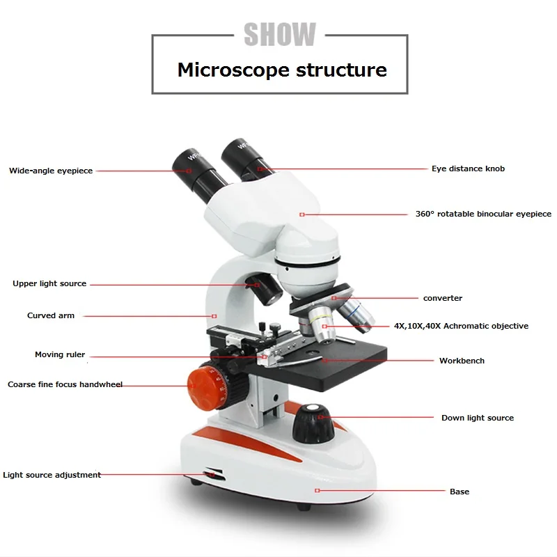
Mikroskop Binokular Biologi 40-5000X Set Portabel Percobaan Deteksi Ilmiah Perbesaran Tinggi Definisi Tinggi
Simple Microscope - Diagram (Parts labelled), Principle, Formula and Uses Optical microscope - These use transparent lenses and visible light to see objects to the order of one micrometer ( one millionth of one meter Charged Particle (ion and electron) microscope - These microscopes employ electrostatic or electromagnetic lens along with a beam of charged particles to focus on specimens. These microscopes can see objects as tiny as one nanometer ( one tenth billionth of one meter)
Parts of the Microscope with Labeling (also Free Printouts) Parts of the Microscope with Labeling (also Free Printouts) A microscope is one of the invaluable tools in the laboratory setting. It is used to observe things that cannot be seen by the naked eye. Table of Contents 1. Eyepiece 2. Body tube/Head 3. Turret/Nose piece 4. Objective lenses 5. Knobs (fine and coarse) 6. Stage and stage clips 7. Aperture
Microscope Labeling - The Biology Corner This simple worksheet pairs with a lesson on the light microscope, where beginning biology students learn the parts of the light microscope and the steps needed to focus a slide under high power. The labeling worksheet could be used as a quiz or as part of direct instruction where students label the microscope as you go over what each part is used for.
High-resolution holotomography with incoherent light source Tomocube has launched an optical microscope that uses incoherent light to generate holographic images of unlabelled live cells.. The HT-X1 is said to be ideally suited to higher-throughput and automated screening applications, thanks to its ability to image multi-well plate formats, large field-of-view, laser autofocus system, and a very high performance 0.95NA objective.
Microscope Parts and Functions The specimen is placed on the glass and a cover slip is placed over the specimen. This allows the slide to be easily inserted or removed from the microscope. It also allows the specimen to be labeled, transported, and stored without damage. Stage: The flat platform where the slide is placed.
A Study of the Microscope and its Functions With a Labeled Diagram These labeled microscope diagrams and the functions of its various parts, attempt to simplify the microscope for you. However, as the saying goes, 'practice makes perfect', here is a blank compound microscope diagram and blank electron microscope diagram to label.
Required practical - using a light microscope - BBC Bitesize Record the microscope images using labelled diagrams or produce digital images. When first examining cells or tissues with low power, draw an image at this stage, even if going on to examine the...
› cemf › whatisemWhat is Electron Microscopy? - UMASS Medical School Conventional scanning electron microscopy depends on the emission of secondary electrons from the surface of a specimen. Because of its great depth of focus, a scanning electron microscope is the EM analog of a stereo light microscope. It provides detailed images of the surfaces of cells and whole organisms that are not possible by TEM.















Post a Comment for "44 light microscope with labels"