44 labeled compound microscope
Compound Microscope Labeled Diagram | Quizlet Compound Microscope Labeled + − Flashcards Learn Test Match Created by meganplocher734 Terms in this set (14) Eyepiece/Ocular lens Contains the ocular lens Body tube A hollow cylinder that holds the eyepiece. Arm Part that supports the microscope. Stage Supports the slide or specimen Coarse adjustment Knob Parts of a Compound Microscope and Their Functions - NotesHippo Compound microscope magnification is determined by multiplying the eyepiece and objective powers. When viewed through a 5X eyepiece with a 10X objective, an item is magnified 5 x 10=50 times. The magnification is 10 x 45 = 450 times when using a 10X eyepiece and a 45X objective. How to Use the Compound Microscope
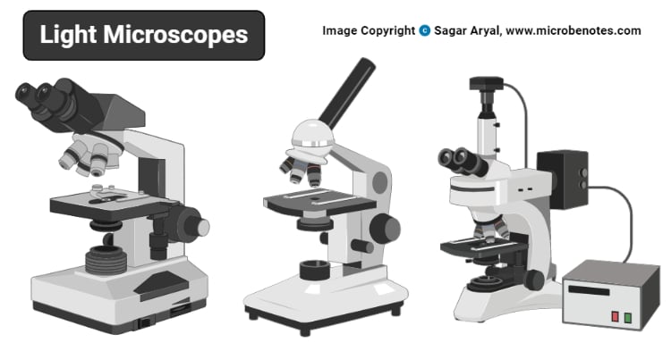
What is a Compound Microscope? | Microscope World Blog A compound microscope is an upright microscope that uses two sets of lenses (a compound lens system) to obtain higher magnification than a stereo microscope. A compound microscope provides a two-dimensional image, while a stereo microscope provides a three-dimensional image. Compound microscopes typically provide magnification in the range of ...

Labeled compound microscope
Microscope Parts, Function, & Labeled Diagram - slidingmotion There are so many individual parts in the microscope. But, categorization of the compound microscope parts takes place in structural & optical parts. We are going to see these all parts & their functions in detail. Head. The Head is a part of a microscope that is on the upper side of the microscope and carries an optical lens. Base Parts of a microscope with functions and labeled diagram 17.9.2022 · Q. Differentiate between a condenser and an Abbe condenser. Ans. Condensers are lenses that are used to collect and focus light from the illuminator into the specimen. They are found under the stage next to the diaphragm of the microscope. They play a major role in ensuring clear sharp images are produced with a high magnification of 400X and above. Compound Microscope - Types, Parts, Diagram, Functions and Uses Compound microscope - It is an optical instrument consists of two convex lenses of short focal lengths primarily used for observing a highly magnified image of minute objects. Lenses Simple microscope - It has a convex lens. It uses only one lens to magnify objects. An example of a simple microscope is a magnifying glass.
Labeled compound microscope. Compound Microscope- Definition, Labeled Diagram, Principle, … Apr 03, 2022 · Working Principle of the Compound Microscope Compound microscopes have a combination of lenses that enhances both magnifying powers as well as the resolving power. The specimen or object, to be examined is usually mounted on a transparent glass slide and positioned on the specimen stage between the condenser lens and objective lens. compound microscope parts (labeling) Flashcards | Quizlet Study with Quizlet and memorize flashcards containing terms like eyepiece tube - connects the eyepiece to the objective lens, nosepiece (turret) - holds and spins the objective lenses, 4x objective lens - the 'scanning" objective lens with the lowest magnification and more. What is a Compound Microscope? - New York Microscope Company A compound microscope is an instrument that is used to view magnified images of small specimens on a glass slide. It can achieve higher levels of magnification than stereo or other low power microscopes and reduce chromatic aberration. It achieves this through the use of two or more lenses in the objective and the eyepiece. Compound Microscope - Diagram (Parts labelled), Principle and Uses Also called as binocular microscope or compound light microscope, it is a remarkable magnification tool that employs a combination of lenses to magnify the image of a sample that is not visible to the naked eye. Compound microscopes find most use in cases where the magnification required is of the higher order (40 - 1000x).
Microscope Parts and Functions Learning to use and adjust your compound microscope is the next important step. It's also imperative to know and understand the best practices of cleaning your microscope . The microscope parts work together in hospitals and in forensic labs, for scientists and students, bacteriologists and biologists so that they may view bacteria, plant and animal cells and … Parts of Stereo Microscope (Dissecting microscope) – labeled … Stereo microscopes (also called Dissecting microscope) are branched out from other light microscopes for the application of viewing "3D" objects. These include substantial specimens, such as insects, feathers, leaves, rocks, sand grains, gems, coins, and stamps, etc. Functionally, a stereo microscope is like a powerful magnifying glass. Microscope Labeling Game - PurposeGames.com About this Quiz. This is an online quiz called Microscope Labeling Game. There is a printable worksheet available for download here so you can take the quiz with pen and paper. This quiz has tags. Click on the tags below to find other quizzes on the same subject. Science. Brightfield Microscope (Compound Light Microscope)- Definition ... 15.4.2022 · Compound Microscope- Definition, Labeled Diagram, Principle, Parts, Uses; Disadvantages of Brightfield microscope. The aperture diaphragm may cause great contrast which may distort the outcome of the image, therefore iris diaphragm is preferred.
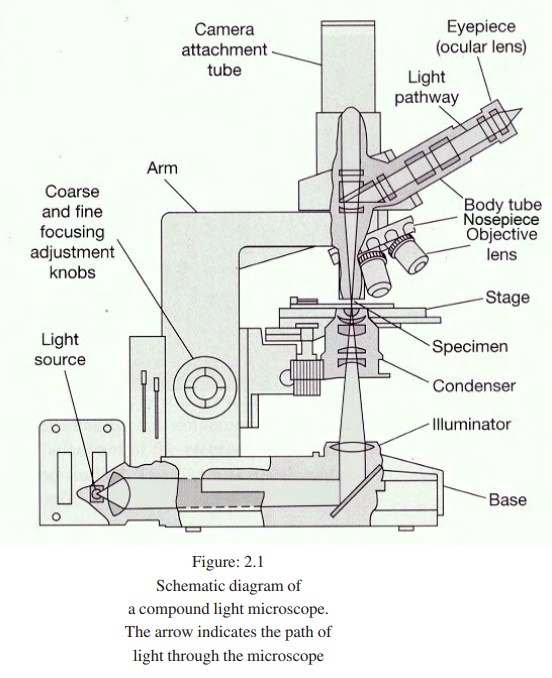
Light Microscope- Definition, Principle, Types, Parts, Labeled … 7.9.2022 · Brightfield Light Microscope (Compound light microscope) This is the most basic optical Microscope used in microbiology laboratories which produces a dark image against a bright background. Made up of two lenses, it is widely used to view plant and animal cell organelles including some parasites such as Paramecium after staining with basic stains. UD Virtual Compound Microscope - University of Delaware ©University of Delaware. This work is licensed under a Creative Commons Attribution-NonCommercial-NoDerivs 2.5 License.Creative Commons Attribution-NonCommercial-NoDerivs 2 A Study of the Microscope and its Functions With a Labeled Diagram ... To better understand the structure and function of a microscope, we need to take a look at the labeled microscope diagrams of the compound and electron microscope. These diagrams clearly explain the functioning of the microscopes along with their respective parts. Man's curiosity has led to great inventions. The microscope is one of them. Parts of the Microscope with Labeling (also Free Printouts) Parts of the Microscope with Labeling (also Free Printouts) By Editorial Team March 7, 2022 A microscope is one of the invaluable tools in the laboratory setting. It is used to observe things that cannot be seen by the naked eye. Table of Contents 1. Eyepiece 2. Body tube/Head 3. Turret/Nose piece 4. Objective lenses 5. Knobs (fine and coarse) 6.
This is a common compound microscope. Label its parts from A ...
Lappeenranta - South Karelia Museum - Lappeenrannan kaupunki Kristiinankatu 15, The Fortress of Lappeenranta Tel. +358 5 616 2255 Tel. +358 40 587 2274 . Opening hours OPENING HOURS. Winter season: 1 Jan-5 June and 22 Aug-30 Dec

Compound Microscope
Label the microscope — Science Learning Hub All microscopes share features in common. In this interactive, you can label the different parts of a microscope. Use this with the Microscope parts activity to help students identify and label the main parts of a microscope and then describe their functions. Drag and drop the text labels onto the microscope diagram.

A Study of the Microscope and its Functions With a Labeled ...
Microscope Types (with labeled diagrams) and Functions Compound microscope labeled diagram Compound microscope functions: It finds great application in areas of pathology, pedology, forensics etc Its greater order of magnification allows for deeper study of microbial organisms to Detect the cause of diseases Study the mineral composition in soils

Compound Microscope- Definition, Labeled Diagram, Principle ...
16 Parts of a Compound Microscope: Diagrams and Video In compound microscopes with two eye pieces there are prisms contained in the body that will also split the beam of light to enable you to view the image through both eye pieces. 2. Arm. The arm of the microscope is another structural piece. The arm connects the base of the microscope to the head/body of the microscope.

The Parts of a Compound Microscope and How To Handle Them ...
Compound Microscope: Parts of Compound Microscope - BYJUS (A) Mechanical Parts of a Compound Microscope 1. Foot or base It is a U-shaped structure and supports the entire weight of the compound microscope. 2. Pillar It is a vertical projection. This stands by resting on the base and supports the stage. 3. Arm The entire microscope is handled by a strong and curved structure known as the arm. 4. Stage

Microscope With Labels clip art | Microscope parts ...
Microscope, Microscope Parts, Labeled Diagram, and Functions 3.9.2022 · Revolving Nosepiece or Turret: Turret is the part of the microscope that holds two or multiple objective lenses and helps to rotate objective lenses and also helps to easily change power. Objective Lenses: Three are 3 or 4 objective lenses on a microscope. The objective lenses almost always consist of 4x, 10x, 40x and 100x powers. The most common eyepiece lens is …

Microscopes: A Beginner's Guide
Compound Microscope Parts – Labeled Diagram and their … Major structural parts of a compound microscope. There are three major structural parts of a compound microscope. The head includes the upper part of the microscope, which houses the most critical optical components, and the eyepiece tube of the microscope.; The base acts as the foundation of microscopes and houses the illuminator.; The arm connects between the base …
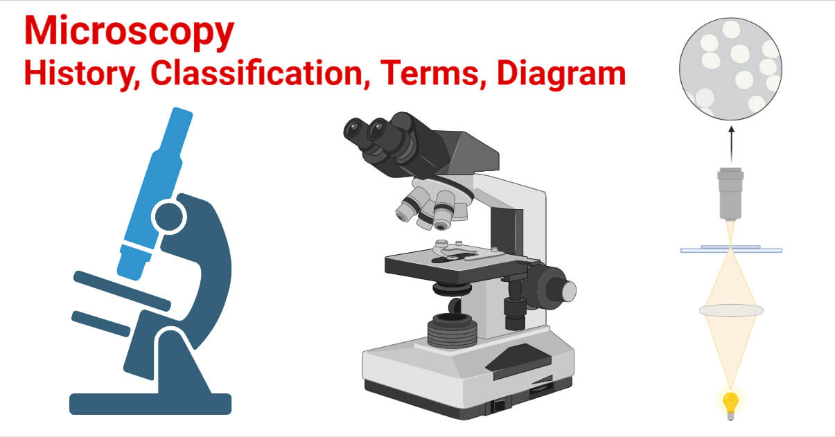
Microscopy- History, Classification, Terms, Diagram
Compound Microscope Parts Compound Microscope Parts A high power or compound microscope achieves higher levels of magnification than a stereo or low power microscope. It is used to view smaller specimens such as cell structures which cannot be seen at lower levels of magnification. Essentially, a compound microscope consists of structural and optical components.

Parts of the Microscope with Labeling (also Free Printouts ...
Why Is The Light Microscope Called A Compound Microscope? A typical compound microscope will allow you a magnification that will go from 40x to 1000x, from which you can easily observe the bacteria. This higher magnification is produced when the compound microscope uses to lens to magnify instead of using just one.

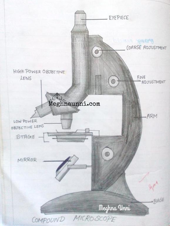
How to draw compound of Microscope easily - step by step
Labelled Diagram of Compound Microscope The below mentioned article provides a labelled diagram of compound microscope. Part # 1. The Stand: The stand is made up of a heavy foot which carries a curved inclinable limb or arm bearing the body tube. The foot is generally horse shoe-shaped structure (Fig. 2) which rests on table top or any other surface on which the microscope in kept.
MICROBIO 16 Parts of a Compound Microscope with Diagram and ...
How to Use a Compound Microscope Grasp the arm with one hand and place the other hand under the base for support. Always place the microscope on a level and stable surface. Slide Preparation: Microscope slides should always be prepared with a cover slip or cover glass over the specimen. This will help protect the objective lenses if they touch the slide.

Compound Microscope Parts – Labeled Diagram and their ...
Compound Light Microscope: Everything You Need to Know A compound microscope is a type of light microscope that uses a compound lens system to magnify specimens for up to 1000x or more. It's made up of at least one objective lens and at least one ocular lens, as well as a light source, condenser, and other essential parts.

Spongelab | Explore | Masterlist
Compound Microscope Parts, Functions, and Labeled Diagram Nov 18, 2020 · Parts of a Compound Microscope Each part of the compound microscope serves its own unique function, with each being important to the function of the scope as a whole. The individual parts of a compound microscope can vary heavily depending on the configuration & applications that the scope is being used for. Common compound microscope parts include: Compound Microscope Definitions for ...
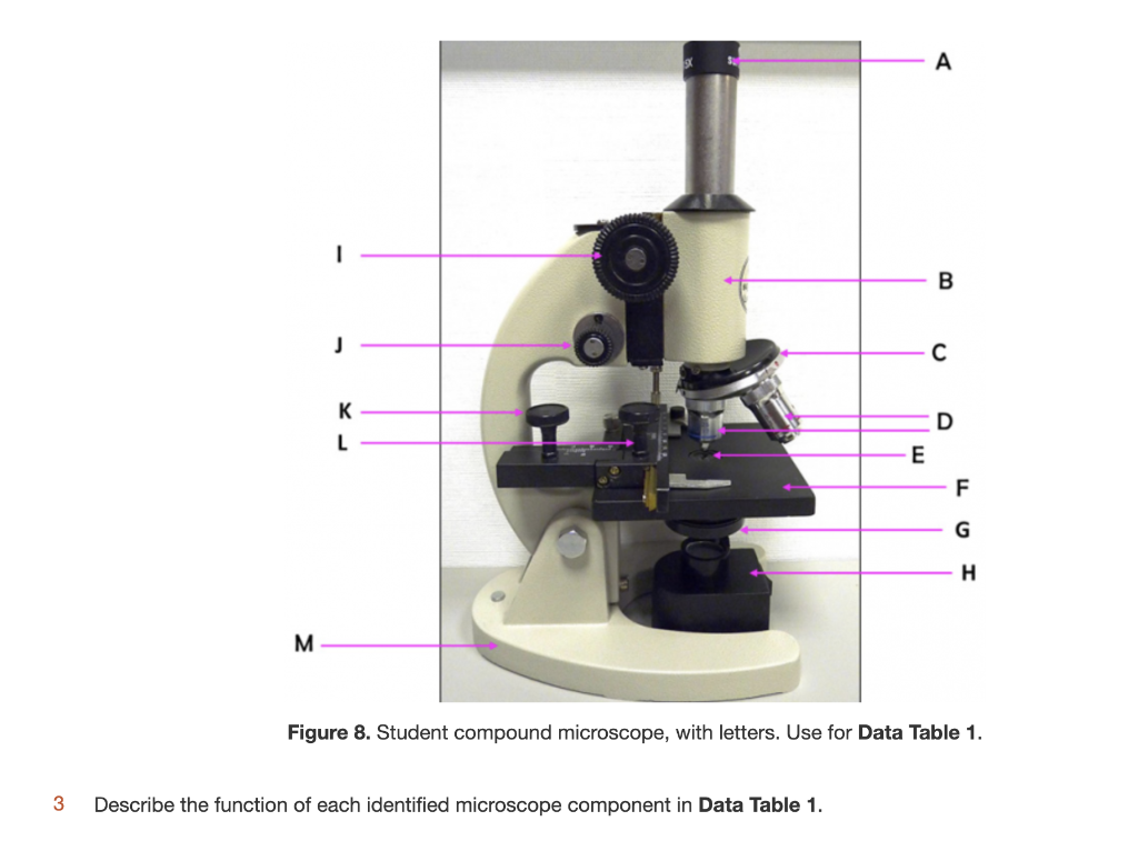
Solved - B J с K D L E F G H M Figure 8. Student compound ...
Compound Microscope: Definition, Diagram, Parts, Uses, Working ... - BYJUS The compound microscope is mainly used for studying the structural details of cell, tissue, or sections of organs. The parts of a compound microscope can be classified into two: Non-optical parts Optical parts Non-optical parts Base The base is also known as the foot which is either U or horseshoe-shaped.

Simple Microscope - Diagram (Parts labelled), Principle ...
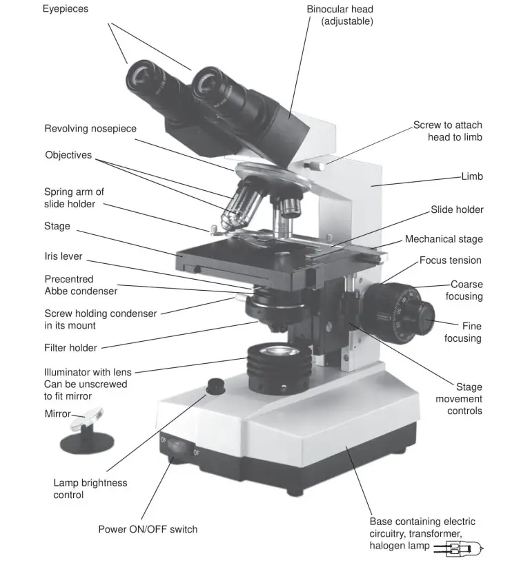
Binocular Microscope Anatomy - Parts and Functions with a Labeled ... Now, I will discuss the details anatomy of the light compound microscope with the labeled diagram. Why it is called binocular: because it has two ocular lenses or an eyepiece on the head that attaches to the objective lens, this ocular lens magnifies the image produced by the objective lens. Binocular microscope parts and functions

Solved A. OLYMPUS C. B. Use the Diagram to answer the | Chegg.com
Microscope Labeling - The Biology Corner Microscope Labeling. Shannan Muskopf May 31, 2018. This simple worksheet pairs with a lesson on the light microscope, where beginning biology students learn the parts of the light microscope and the steps needed to focus a slide under high power. The labeling worksheet could be used as a quiz or as part of direct instruction where students ...

Cytology. Cytology. radiation used to illuminate the specimen ...
Compound Microscope - Types, Parts, Diagram, Functions and Uses Compound microscope - It is an optical instrument consists of two convex lenses of short focal lengths primarily used for observing a highly magnified image of minute objects. Lenses Simple microscope - It has a convex lens. It uses only one lens to magnify objects. An example of a simple microscope is a magnifying glass.

Can someone can send me diagram of this compound microscope ...
Parts of a microscope with functions and labeled diagram 17.9.2022 · Q. Differentiate between a condenser and an Abbe condenser. Ans. Condensers are lenses that are used to collect and focus light from the illuminator into the specimen. They are found under the stage next to the diaphragm of the microscope. They play a major role in ensuring clear sharp images are produced with a high magnification of 400X and above.
The Compound Light Microscope Label the following parts on ...
Microscope Parts, Function, & Labeled Diagram - slidingmotion There are so many individual parts in the microscope. But, categorization of the compound microscope parts takes place in structural & optical parts. We are going to see these all parts & their functions in detail. Head. The Head is a part of a microscope that is on the upper side of the microscope and carries an optical lens. Base
Compound Microscope Parts, Diagram Definition, Application ...

Labeling the Parts of the Microscope | Microscope activity ...

Compound Microscope Parts, Functions, and Labeled Diagram ...

Compound Microscope Parts – Labeled Diagram and their ...
Biology : Compound Microscope Diagram for Class 8 ...
The compound light microscope

Compound Microscope Parts – Labeled Diagram and their ...

Draw a neat labelled diagram of a compound microscope class ...

Compound Microscope Parts, Function, & Diagram | What is a ...

Parts of a microscope with functions and labeled diagram
Compound Microscope - Types, Parts, Diagram, Functions and ...

Labeling the Parts of the Microscope | Microscope World Resources

2.1 " Compound Microscope" | Download Scientific Diagram

Label the microscope — Science Learning Hub

Microscope Labeling
Parts of a Microscope with Their Functions – Microbe Online

Solved Lab 2: Use and Care of the Light Compound Microscope ...

Compound Microscope: Know Definition,working, diagram, properties

Label Microscope Diagram - EnchantedLearning.com
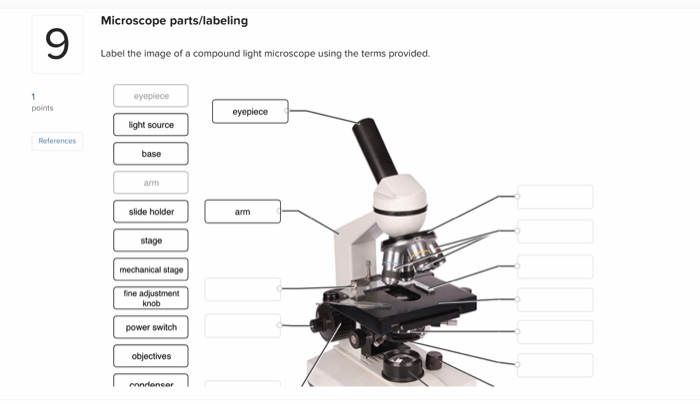
Solved Microscope parts/labeling 9 Label the image of a ...

Compound microscope - their parts and function - Microscopy4kids

Microscope Parts & Specifications | Microscope parts, Health ...

compound microscope Diagram | Quizlet

Compound Microscope Parts – Labeled Diagram and their ...
Light Microscope- Definition, Principle, Types, Parts ...
Post a Comment for "44 labeled compound microscope"