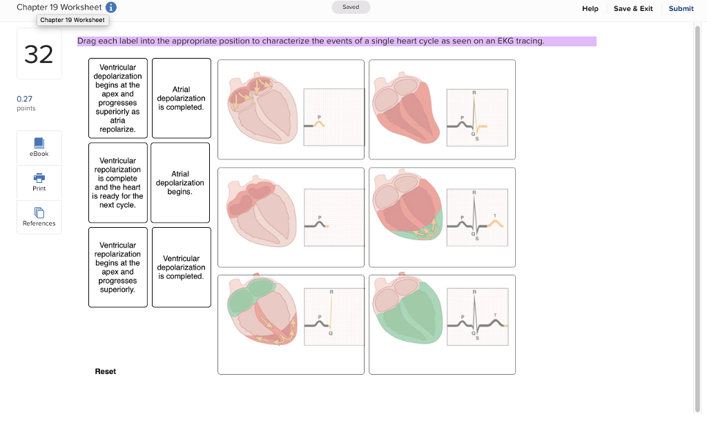
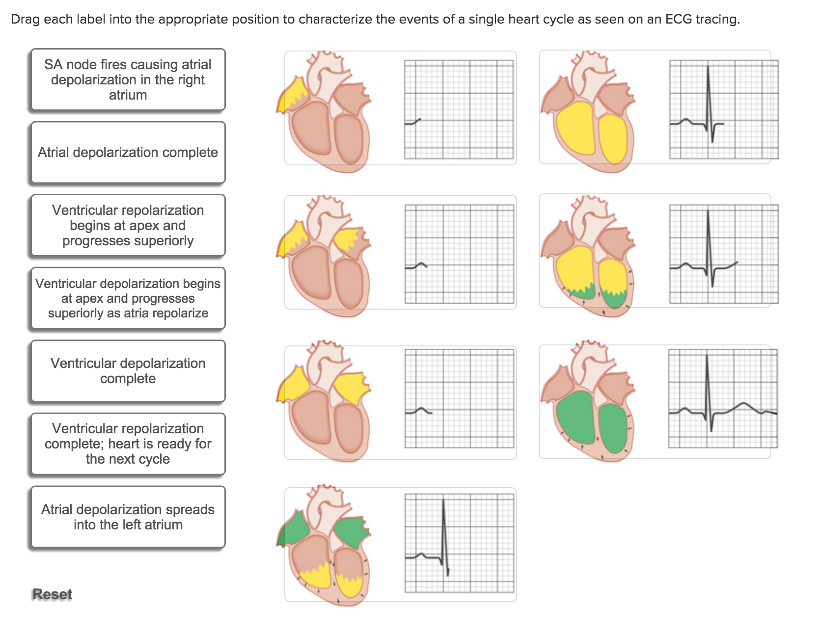
39 drag each label into the appropriate position to characterize the events of a single heart cycle as seen on an ecg tracing.
Ch 20 - Edited Cardiac Cycle, Part 2.pdf - Course Hero There are many ways to describe a single heartbeat or cardiac cycle. Examine the model and as a group define a cardiac cycle. When you are prompted, the instructor will have the reporter for each group write your response on the board. The reporter should be able to verbally explain your definition. Chapter 19,20,21 Flashcards | Quizlet Drag each label into the appropriate position to characterize the events of a single heart cycle as seen on an ECG tracing. Drag each statement to the appropriate position to identify the valve being described. The __________ valve is between the right atrium and right ventricle. Tricuspid Select all that are true regarding ventricular balance. 2,4
Solved Drag each label into the appropriate position to - Chegg Drag each label into the appropriate position to characterize the events of a single heart cycle as seen on an ECG tracing. Show transcribed image text Expert Answer 100% (22 ratings) Figure 1 SA node fires causing atrial depolarization in right atrium Figure 5 ventricular de … View the full answer

Drag each label into the appropriate position to characterize the events of a single heart cycle as seen on an ecg tracing.
Human Anatomy & Physiology Laboratory Manual Main Version 10th Edition ... If an incision cuts the heart into right and left parts, the section is a 15 section; but if the heart is cut so that superior and inferior portions result, the section is a 16 section. You are told to cut a dissection animal along two planes so that both kidneys are observable in each section. Solved Drag each label into the appropriate position to - Chegg Transcribed image text: Drag each label into the appropriate position to characterize the events of a single heart cycle as seen on an EKG tracing. Ventricular repolarization begins at the apex and progresses superiorly Ventricular repolarization is complete and the heart is ready for the next cycle Ventricular depolarization is completed Ventricular depolarization begins at the apex and ... ReaderUi ReaderUi
Drag each label into the appropriate position to characterize the events of a single heart cycle as seen on an ecg tracing.. BIOLOGY K - Essay Help Ch 56-58 i Saved He O Indicate whether each label describes organisms at the r-selected end or the K-selected end of the continuum Life history strategies differ along a continuum from what is referre… ASAp Please! 1. You are working with animal cells that have an internal (cytosol) solute concentration of 5 mM solute. Solved Drag each label into the appropriate position to | Chegg.com 83% (6 ratings) Parts of Normal ECG: 1. P wave: It denotes atrial depolarization. 2. QRS Complex: This is caused by ventricular depolarization. 3. Q wave: If the first wave of …. View the full answer. Transcribed image text: Drag each label into the appropriate position to identify the waves of a normal ECG Alpha wave 0 QRS complex ST segment ... Heart Lecture Flashcards | Quizlet Correctly label the following external anatomy of the posterior heart. Drag each label into the appropriate position to characterize the events of a single heart cycle as seen on an EKG tracing. Correctly label the pathway of blood flow through the heart, beginning with the right atrium. Ch. 19 Circulatory System- heart Flashcards | Quizlet Correctly label the external anatomy of the anterior heart. Place the labels in order denoting the flow of blood through the pulmonary circuit beginning with the right atrium and ending in the left atrioventricular valve. The first and last structures are given. Right atrium 1. tricuspid valve 2. right ventricle 3. pulmonary valve
BILOGY C - Essay Help Use the following terms and label the life cycle shown in Figure 6-2 on the next page. In the table below, indicate the ploidy and function. On the diagram, also indicate where meiosis and syngamy ta… Ericka was supposed to tell a certain guy that her best friend had a crush on him. Instead, she ended up hooking up with the guy. Chapter 15 Cardiovascular Practice Flashcards & Practice Test - Quizlet Complete each sentence, and then place them in the correct order to describe blood flow through the heart, beginning with blood entering the right side of the heart. Beginning with the return from the systemic circulation, blood enters the right atrium. Blood then travels through the tricuspid valve and into the right ventricle. Respiratory Anatomy - YouTube About Press Copyright Contact us Creators Advertise Developers Terms Privacy Policy & Safety How YouTube works Test new features Press Copyright Contact us Creators ... Cardiac Muscle and Electrical Activity - Lumen Learning Normal cardiac rhythm is established by the sinoatrial (SA) node, a specialized clump of myocardial conducting cells located in the superior and posterior walls of the right atrium in close proximity to the orifice of the superior vena cava. The SA node has the highest inherent rate of depolarization and is known as the pacemaker of the heart.
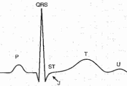
Emergency Care - PMC Central venous pressure refers to the hydrostatic pressure in the anterior vena cava and is influenced by vascular fluid volume, vascular tone, function of the right side of the heart, and changes in intrathoracic pressure during the respiratory cycle. The CVP is not a true measure of blood volume but is used to gauge fluid therapy as a method ... Module 3.docx - 1. Describe in your own words what each... The ST segment is the flat line on the ECG; end of the S wave and beginning of the T wave. This segment is when ventricular depolarization and repolarization occurs. Mechanisms that may cause this to change may be due to different infections, such as Pericarditis or other abnormalities. Maternal & Newborn Health Pharmacology Flashcards by RICA ... - Brainscape The Trendelenburg position makes use of gravity to pull the embryo back into the uterus, relieving pressure off the umbilical cord from the presenting part. Cord prolapse is an obstetric emergency. The nurse should suspect it if fetal bradycardia or variable decelerations occur especially, immediately after the rupture of membranes. BIO 211 : anatomy and physiology - Horry-Georgetown Technical College Physiology is the study of how the body of an organism functions. Hole's Human Anatomy & Physiology Ch 3, Section 3.1 Cells Are the Basic Units of the Body, Exercise 1 While very small, millimeters can be further divided into what used to be called microns. Hole's Human Anatomy & Physiology Ch 4, Section 4.1 Metabolic Processes, Exercise 1
(PDF) SHORT QUESTION AND ANSWERS - Academia.edu Enter the email address you signed up with and we'll email you a reset link.
Question 5 1 1 point which of the following - Course Hero Drag each label into the appropriate position to characterize the events of a single heart cycle as seen on an ECG tracing. Drag each label into the appropriate position to characterize the events of. Q&A. Study on the go. Download the iOS
Chapter 19: The Heart Flashcards - Quizlet -left heart—body—right heart supplies blood to all organs of the body Heart Location In the thoracic cavity, between the lungs in the mediastinum Size, Shape and Position of the Heart •Located in thoracic cavity -specifically in the mediastinum •area between lungs -superior to diaphragm -posterior to sternum
Correlations of limb kinematics and bone strain in frogs and toads Enter the email address you signed up with and we'll email you a reset link.
diagnostic - DocShare.tips Forssmann inserted a small catheter into his antecubital vein, walked to the radiology suite, and took an X-ray of the catheter's position in his right atrium (1). Now a designated landmark in medical history, this revolutionary event demonstrated that the heart could be safely accessed
Solved Drag each label into the appropriate position to | Chegg.com transcribed image text: chapter 19 worksheet g seved help save & exit submit chapter 19 worksheet drag each label into the appropriate position to characterize the events of a single heart cycle as seen on an ekg tracing 2 ventricular begins at the atrial apex and 0.27 points superiorly ass completed repolarize ventricular repolarization and the …
Statistics Questions & Answers in 2022 - Essay Help Each observation corresponds to one purchase occasion. Consider the density-based subspace clustering. The size of a subspace is defined to be the total number of dimensions for this subspace; This project requires you to implement a cycle-by-cycle simulator for an in-order APEX pipeline with 7 pipeline stages, each with a delay of one cycle
Emergency Care - PMC There are many indications for administering transfusions of whole blood and component blood products. Take a stepwise approach for every patient that may require a transfusion. If a patient is at risk for blood loss or is anemic, consider a transfusion. Make a decision on the type of transfusion therapy appropriate for each particular patient.
Eastern Kentucky University Eastern Kentucky University's largest and most comprehensive campaign has exceeded its initial $50 million goal. Although the goal has been met, fundraising efforts continue as part of the Make No Little Plans campaign, supporting student success, academic excellence, and campus revitalization. Comprehensive Campaign Information Give Now.
ReaderUi ReaderUi
Solved Drag each label into the appropriate position to - Chegg Transcribed image text: Drag each label into the appropriate position to characterize the events of a single heart cycle as seen on an EKG tracing. Ventricular repolarization begins at the apex and progresses superiorly Ventricular repolarization is complete and the heart is ready for the next cycle Ventricular depolarization is completed Ventricular depolarization begins at the apex and ...
Human Anatomy & Physiology Laboratory Manual Main Version 10th Edition ... If an incision cuts the heart into right and left parts, the section is a 15 section; but if the heart is cut so that superior and inferior portions result, the section is a 16 section. You are told to cut a dissection animal along two planes so that both kidneys are observable in each section.









Post a Comment for "39 drag each label into the appropriate position to characterize the events of a single heart cycle as seen on an ecg tracing."