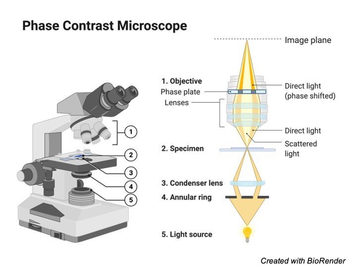
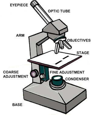
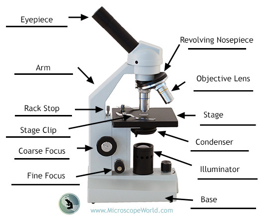
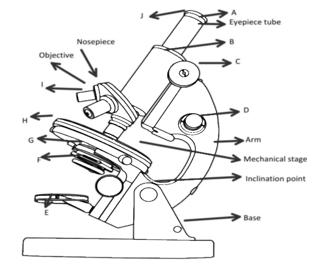
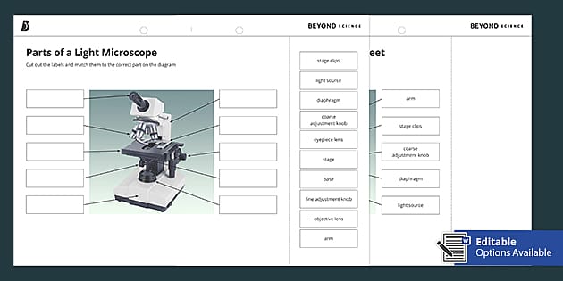
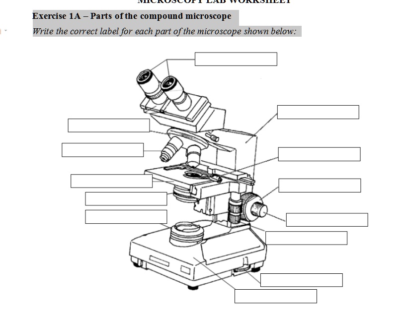
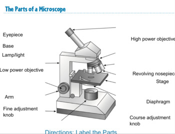
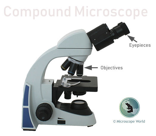
42 microscope parts label
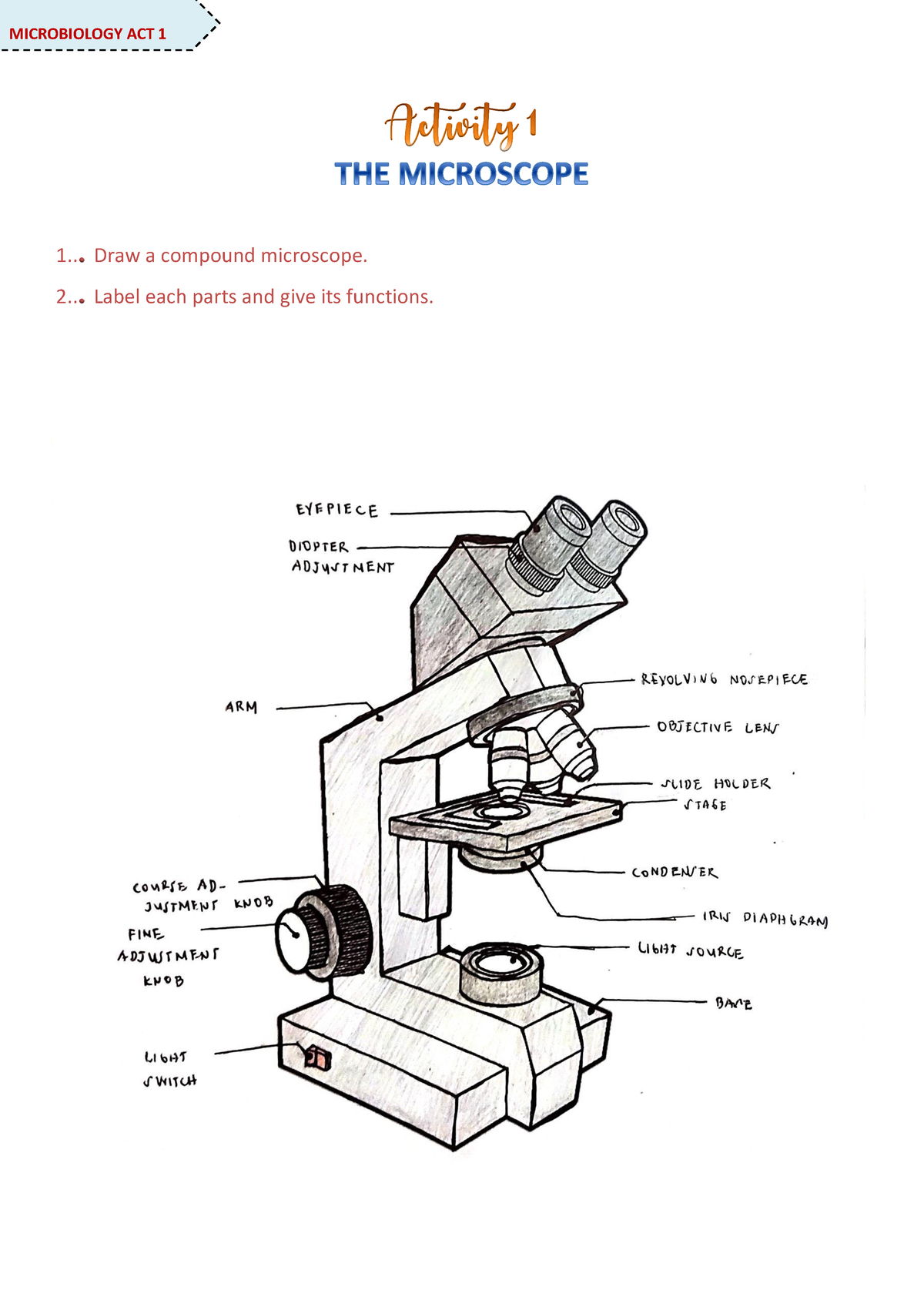
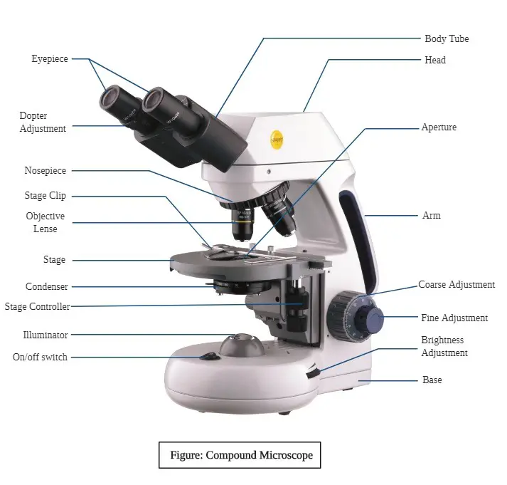
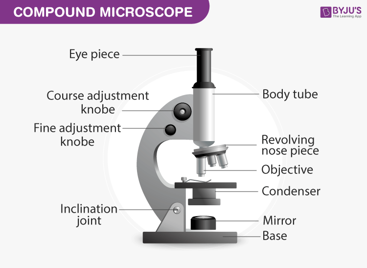
Welcome to Virtual Urchin - University of Washington microscope compare. specimen compare. development & embryology. fertilization lab. embryogenesis to hatching. analyzing gene function. ecology & environment. our acidifying ocean . predator & prey. surfing to settlement. basic biology. urchin anatomy. about us. teacher resources. useful links. Select Language: Welcome to the new Virtual Urchin website! Major … Compound Microscope Parts, Functions, and Labeled Diagram Nov 18, 2020 ... Parts of a Compound Microscope · Eyepiece (ocular lens) with or without Pointer: The part that is looked through at the top of the compound ...
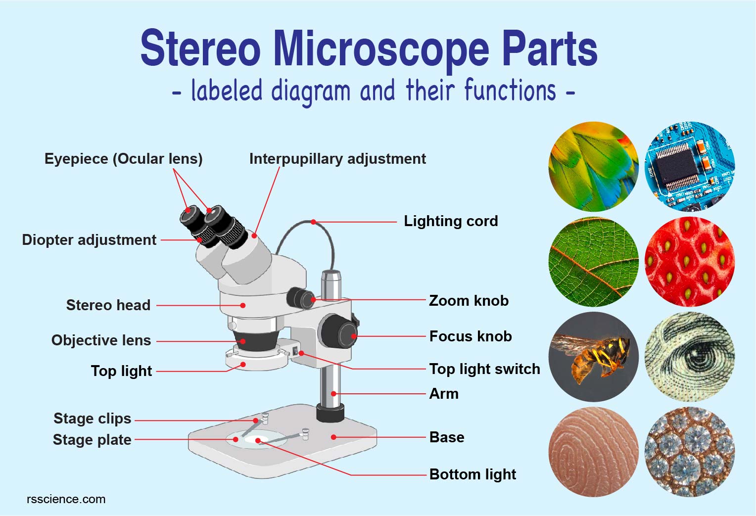
rsscience.com › stereo-microscopeParts of Stereo Microscope (Dissecting microscope) – labeled ... Unlike a compound microscope that offers a flat image, stereo microscopes give the viewer a 3-dimensional image that you can see the texture of a larger specimen. [In this image] Examples of Stereo & Dissecting microscopes. Major microscope brands (Zeiss, Olympus, Nikon, Amscope, Omano, Leica …) all produce stereomicroscopes.

Microscope parts label
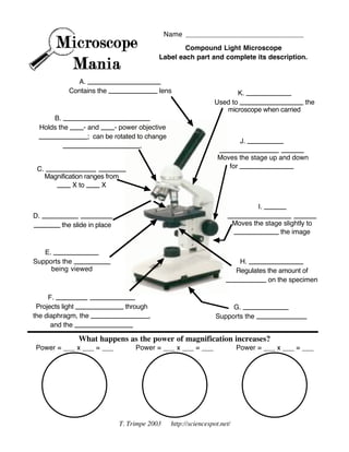
Microscope With Labels clip art - Pinterest Microscope Diagram Labeled, Unlabeled and Blank | Parts of a Microscope. Print a microscope diagram, microscope worksheet, or practice microscope quiz in order ... microbenotes.com › parts-of-a-microscopeParts of a microscope with functions and labeled diagram Sep 17, 2022 · Figure: Diagram of parts of a microscope. There are three structural parts of the microscope i.e. head, base, and arm. Head – This is also known as the body. It carries the optical parts in the upper part of the microscope. Base – It acts as microscopes support. It also carries microscopic illuminators. Parts of Stereo Microscope (Dissecting microscope) – labeled … Optical parts of a stereo microscope work together to magnify and produce a 3-D image of the specimens. These parts include: Eyepieces. The eyepiece (or ocular lens) is the lens part at the top of a microscope that the viewer looks through. Typically, standard eyepieces for a dissecting microscope have a magnifying power of 10x. Optional eyepieces of varying powers are …
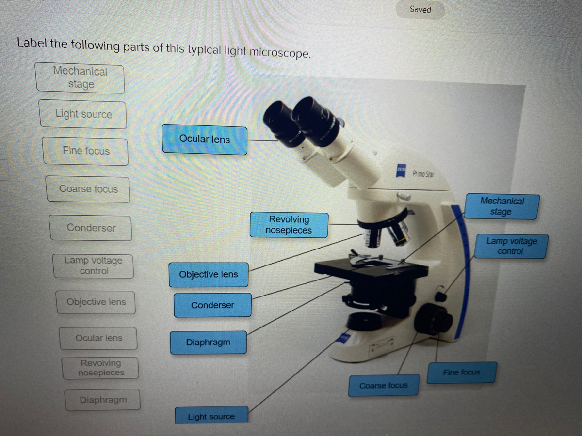
Microscope parts label. Labeling the Parts of the Microscope Labeling the Parts of the Microscope. This activity has been designed for use in homes and schools. Each microscope layout (both blank and the version with ... Virtual Labs: Using the Microscope - GameUp - BrainPOP. In this free online science interactive, students learn the procedures for operating a compound optical light microscope as they would use in a science lab. bVX0-zncj9qJ3G1_r18rkIpQL02X-Oi6tWViR4g4-vwDVmU50WZA-4bRZMjM2TXmc88PAkJ1g0jIembnEbM Compound Microscope Parts – Labeled Diagram and their Functions Compound Microscope Parts – Labeled Diagram and their Functions · Eyepiece · Eyepiece tube · Objective lenses · Nosepiece · Specimen stage · Coarse and fine focus ... UD Virtual Compound Microscope - University of Delaware ©University of Delaware. This work is licensed under a Creative Commons Attribution-NonCommercial-NoDerivs 2.5 License.Creative Commons Attribution-NonCommercial-NoDerivs 2.5 License.
Microscope, Microscope Parts, Labeled Diagram, and Functions Sep 3, 2022 ... Head/Body: It contain the optical parts in the upper part of the microscope. Arm: It supports the tube and connects it to the base. Base: The ... › game › microscope-labelingMicroscope Labeling Game - PurposeGames.com This is an online quiz called Microscope Labeling Game. There is a printable worksheet available for download here so you can take the quiz with pen and paper. This quiz has tags. Click on the tags below to find other quizzes on the same subject. › iet › microscopeVirtual Microscope - NCBioNetwork.org Lesson Description BioNetwork’s Virtual Microscope is the first fully interactive 3D scope - it’s a great practice tool to prepare you for working in a science lab. Explore topics on usage, care, terminology and then interact with a fully functional, virtual microscope. Microscope Imaging Software | Products | Leica Microsystems 20/08/2021 · Microscope Imaging Software. Microscope imaging software from Leica Microsystems combines microscope, digital camera and accessories into one fully integrated solution. With an intuitive user interface and straightforward navigation, it guides the user through any workflow, whether fast image acquisition or sophisticated expert analysis. A ...
Microscope slide - Wikipedia A microscope slide is a thin flat piece of glass, typically 75 by 26 mm (3 by 1 inches) and about 1 mm thick, used to hold objects for examination under a microscope.Typically the object is mounted (secured) on the slide, and then both are inserted together in the microscope for viewing. This arrangement allows several slide-mounted objects to be quickly inserted and … www1.udel.edu › biology › ketchamUD Virtual Compound Microscope - University of Delaware ©University of Delaware. This work is licensed under a Creative Commons Attribution-NonCommercial-NoDerivs 2.5 License.Creative Commons Attribution-NonCommercial-NoDerivs 2 www1.udel.edu › biology › ketchamMicroscopy Pre-lab Activities - University of Delaware Microscope controls: turn knobs (click and hold on upper or lower portion of knob) throw switches (click and drag) turn dials (click and drag) move levers (click and drag) changes lenses (click and drag on objective housing) select a specimen (click on a slide) Parts of the Microscope with Labeling (also Free Printouts) Mar 7, 2022 ... Parts of the Microscope with Labeling (also Free Printouts) · 1. Eyepiece · 2. Body tube/Head · 3. Turret/Nose piece · 4. Objective lenses · 5. Knobs ...
depts.washington.edu › vurchinWelcome to Virtual Urchin - University of Washington Major update Apr 2021: All of the activities on the site are now mobile compatible !! Computers are still recommended, and tablets are preferable to phones: please read the Notes at the bottom of this page for details on the latest updates, mobile compatibility and general information about using this site.
Virtual Microscope - NCBioNetwork.org Lesson Description BioNetwork’s Virtual Microscope is the first fully interactive 3D scope - it’s a great practice tool to prepare you for working in a science lab. Explore topics on usage, care, terminology and then interact with a fully functional, virtual microscope. When you are ready, challenge your knowledge in the testing section to see what you have learned.
Microscope Labeling Game - PurposeGames.com About this Quiz. This is an online quiz called Microscope Labeling Game. There is a printable worksheet available for download here so you can take the quiz with pen and paper.. This quiz has tags. Click on the tags below to find other quizzes on the same subject.
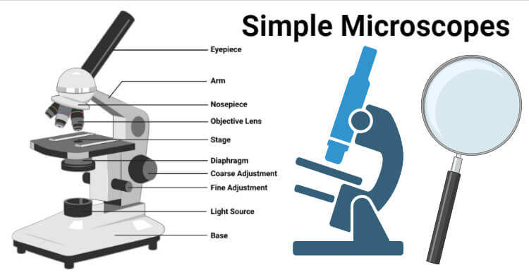
Simple Microscope - Diagram (Parts labelled), Principle, Formula ... Feb 23, 2022 ... The working principle of a simple microscope is that when a lens is held close to the eye, a virtual, magnified and erect image of a specimen is ...
Parts of a microscope with functions and labeled diagram 17/09/2022 · Thank you very much it really helped me with my science home work since i in 8th grade and this my home work to draw a microscope label all the parts and the function thank may the holy father of holy spirits bless you and give more wisdom thanks love you all keep up the good work and thank you again bye. Reply . Sagar Aryal. May 21, 2022 at 1:57 AM . Thank you …
Microscopy Pre-lab Activities - University of Delaware Microscope controls: turn knobs (click and hold on upper or lower portion of knob) throw switches (click and drag) turn dials (click and drag) move levers (click and drag) changes lenses (click and drag on objective housing) select a specimen (click on a slide) adjust oculars (in "through" view, with the light on, click and drag to move oculars closer or further apart) Designed and …
Label the microscope - Science Learning Hub Jun 8, 2018 ... Label the microscope ; base. The bottom of the microscope used for stability ; high-power objective. For increased magnification – usually 10x, ...
Parts of Stereo Microscope (Dissecting microscope) – labeled … Optical parts of a stereo microscope work together to magnify and produce a 3-D image of the specimens. These parts include: Eyepieces. The eyepiece (or ocular lens) is the lens part at the top of a microscope that the viewer looks through. Typically, standard eyepieces for a dissecting microscope have a magnifying power of 10x. Optional eyepieces of varying powers are …
microbenotes.com › parts-of-a-microscopeParts of a microscope with functions and labeled diagram Sep 17, 2022 · Figure: Diagram of parts of a microscope. There are three structural parts of the microscope i.e. head, base, and arm. Head – This is also known as the body. It carries the optical parts in the upper part of the microscope. Base – It acts as microscopes support. It also carries microscopic illuminators.
Microscope With Labels clip art - Pinterest Microscope Diagram Labeled, Unlabeled and Blank | Parts of a Microscope. Print a microscope diagram, microscope worksheet, or practice microscope quiz in order ...
(159).jpg)
















Post a Comment for "42 microscope parts label"