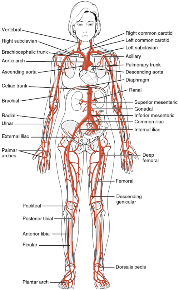
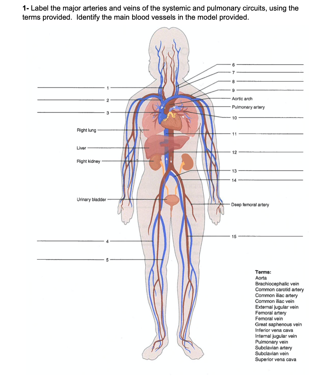
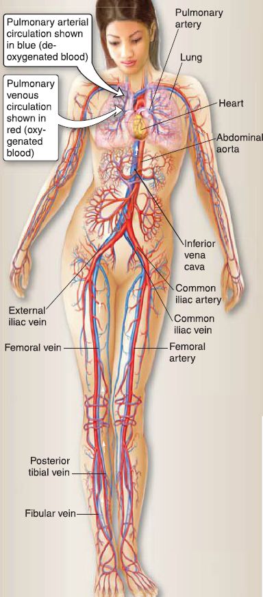
44 label the arteries of systemic circuit.
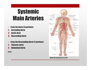
people.ucalgary.ca › ~rosenber › MajorBloodVesselsSYSTEMIC CIRCULATION - University of Calgary in Alberta MAJOR BLOOD VESSELS OF THE SYSTEMIC CIRCULATION OF THE HUMAN BODY ARTERIAL SYSTEM DIVISIONS AND BRANCHES OF THE AORTA (Fig. 14-12) 1. ascending aorta right and left coronary arteries arise as the aorta leaves the heart and carry blood into the coronary circuit. 2. arch of the aorta left and right common carotid arteries subclavian arteries. Understudied Populations in PAH (Transcript) Dr Aboulhosn discusses the role of congential heart defects in pulmonary hypertension, and other understudied populations with PH.
And Answers Worksheet Heart Circulation answers: heart anatomy label the interior of the heart in this printout ) the circulatory system transports to the tissues and organs of the body the oxygen, nutritive substances, immune substances, hormones, and chemicals necessary for normal function and activities of the organs; it also carries ) the circulatory system transports to the …
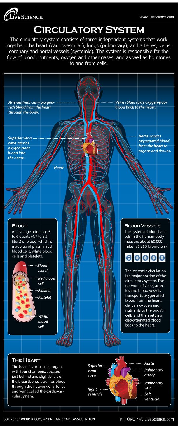
Label the arteries of systemic circuit.
Important Question for Class 10 Science Life Processes - Learn CBSE (a) (i) Unused carbohydrates in plants are stored in the form of complex sugar known as starch. They are later broken down into simple sugars (glucose) when energy is needed. (ii) The assimilated food molecules hold energy in their chemical bonds. Their bond energy is released by oxidation in the cell. › homework-help › questions-andSolved Label the arteries of the systemic circuit. Please ... Transcribed image text: Label the arteries of systemic circuit. Axillary a. Femoral a. Common iliac a. Subclavian a. External iliac a. Brachiocephalic trunk Deep femoral a. Internal iliac a. External iliac a. Popliteal a. Ulnar a. Common carotid a. Aorta Internal iliac a. Brachial a. Radial a. Human Circulatory system | Teaching Resources Distinguish between pulmonary and systemic circuits labels and function of the heart explanation of cardiac cycle explanation of how an impulse is generated in the heart labeling and functions of the major arteries and veins in the circulatory system comparing structure and function of the three main blood vessels
Label the arteries of systemic circuit.. Heart - Wikipedia The heart is a muscular organ in most animals.This organ pumps blood through the blood vessels of the circulatory system. The pumped blood carries oxygen and nutrients to the body, while carrying metabolic waste such as carbon dioxide to the lungs. In humans, the heart is approximately the size of a closed fist and is located between the lungs, in the middle compartment of the chest. quizlet.com › 637928144 › chapter-13-ap-circulatoryChapter 13 A&P circulatoryII-Heart and blood - Quizlet Place the labels in order denoting the flow of blood through the structures of the heart beginning with the vena cava. 1. Superior vena cavae 2. Right atrium 3. Tricuspid valve 4. Right ventricle 5. Pulmonary valve 6. Pulmonary trunk 7. Pulmonary artery 8. Lungs 9. Pulmonary vein 10. Left atrium 11. Bicuspid valve 12. Left ventricle 13. Worksheet Heart The Of Anatomy - teg.bbs.fi.it Search: Anatomy Of The Heart Worksheet. Anatomy of the Heart - Chute Heart Anatomy Location of the Heart Quiz: Heart & Circulatory System The peripheral nervous system includes nerves that go from your central nervous system out to all parts of your body Larger text size ), with a blank version to be filled in and an answer version ), with a blank version to be filled in and an answer version. Coronary arteries and cardiac veins: Anatomy and branches | Kenhub The conus arteriosus branch of the right coronary artery (otherwise called the right conus artery or the arteria coni arteriosi) is the first branch of the right coronary artery. In some instances, it arises directly from the right coronary sinus; at which point it is referred to as the third coronary artery.
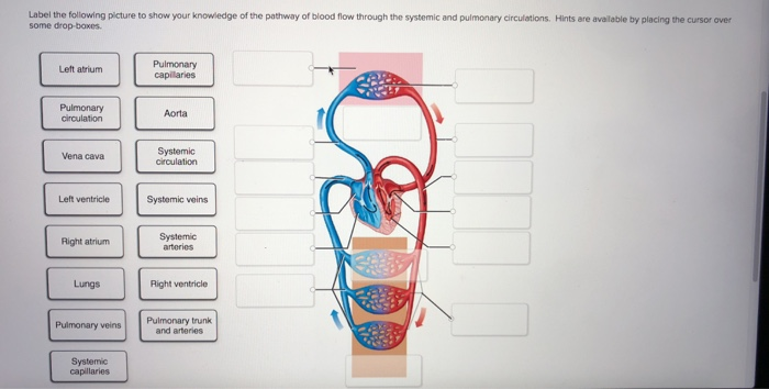
quizlet.com › 501510723 › lab-4-exercises-30-31Lab 4 : Exercises 30 & 31 : Blood vessels Flashcards - Quizlet Label the structures associated with the pulmonary and systemic circuits by clicking and dragging the labels to the correct location. Label the major arteries of the abdominal, mesenteric, and pelvic areas by clicking and dragging the labels to the correct location. Label the arteries of the brain by clicking and dragging the labels to the correct location. quizlet.com › 572157837 › apr-cardiovascular-andAPR Cardiovascular and Blood Flashcards - Quizlet Label the arteries of systemic circuit. Drag and drop each label, indicating whether each artery arises directly from the aorta (Aorta) or from another (Other) artery. Three arteries branch off of the aortic arch. Circulatory system - Wikipedia The circulatory system includes the heart, blood vessels, and blood. The cardiovascular system in all vertebrates, consists of the heart and blood vessels. The circulatory system is further divided into two major circuits - a pulmonary circulation, and a systemic circulation. The pulmonary circulation is a circuit loop from the right heart taking deoxygenated blood to the lungs where it is ... Systolic vs. Diastolic Blood Pressure - Verywell Health During a heartbeat, the heart pushes blood out into the arteries. Systolic pressure is the measure of this force. This phase, known as systole, is the point at which blood pressure is the highest. Systolic blood pressure is considered normal when the reading is below 120 mmHg (millimeters of mercury) while a person is sitting quietly at rest. 1
Of Anatomy Worksheet Heart The this coordinates with our human heart labeling worksheet below understanding the anatomy of the mouth, with information on the teeth and jaw, the gingiva, tongue, palate, cheeks and lips located between the lungs in the middle of the chest, the heart pumps blood through the network of arteries and veins known as the cardiovascular system an … Anatomy Worksheet Of The Heart On average, the heart beats about 100,000 times a day, i Also covered is a full description of how the frog's three-chambered heart works Quizzes by RM Chute Respiration and Excretion System Organs: heart, lung, kidney, ureter, urinary bladder, cloaca Before observing internal or external structures of the fetal pig use your dissection manual textbook and dissection notebook to answer the pre ... Diagram of Human Heart and Blood Circulation in It There are three arteries of the heart, including pulmonary artery, aorta, and coronary arteries. Heart Of The Anatomy Worksheet Search: Anatomy Of The Heart Worksheet. 00 KB] Circulatory Systems Primary Functions -Transport oxygen and nutrients to actively metabolizing tissues, Diffusion, Circulation Time, Pumping Structure, …
Society for Cardiovascular Magnetic Resonance/European Society of ... The ultimate goal of surgery is to completely separate the systemic and pulmonary circulations and place them in a "series circuit." ... (if present), pulmonary stenosis, the pulmonary arteries and aortic to pulmonary collaterals. At the BDG/hemiFontan stage, besides reassessment of the aortic arch, the superior cava connections ...
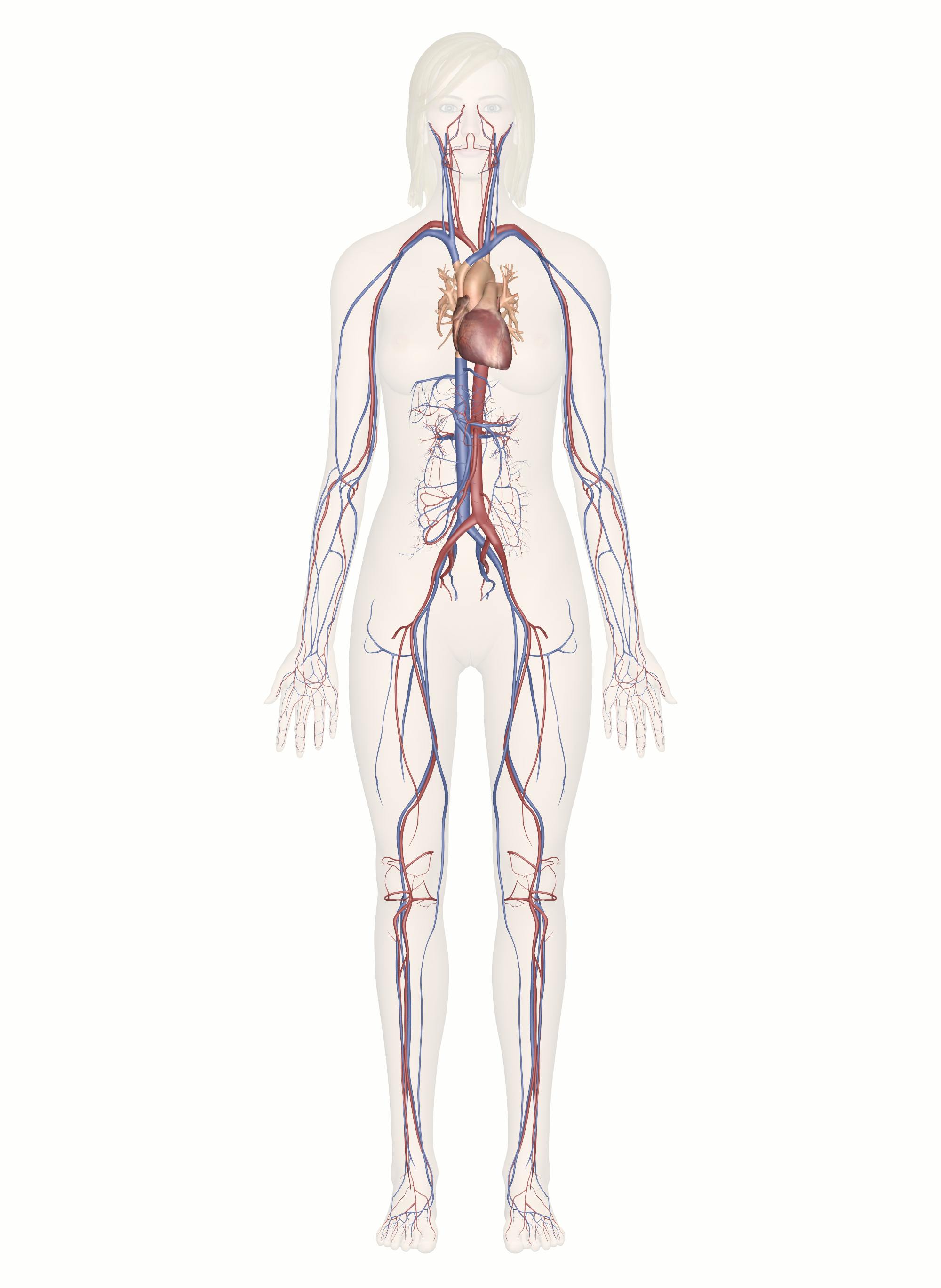
Circulatory System Diagram | New Health Advisor Coronary circuit mainly consists of cardiac veins including anterior cardiac vein, small vein, middle vein and great (large) cardiac vein. There are different types of circulatory system diagrams; some have labels while others don't. The color blue stands for deoxygenated blood while red stands for blood which is oxygenated.
The Musculoskeletal System and Disease - Verywell Health Musculoskeletal is a general term which, as its name suggests, relates to the muscles and the skeleton of the body. More specifically, the musculoskeletal system includes bones, muscles, joints, cartilage, ligaments, tendons, and bursae. The musculoskeletal system provides stability and also allows for movement of the body.
Worksheet Of The Anatomy Heart The Heart, part 1 - Under Pressure: Crash Course A&P #25 The Anatomy And Physiology Printable Worksheets could be printed on regular paper and may be produced use to incorporate all of the extra info about the college students These pages contain worksheets and lessons that are ready for you to print out and work on off-line Learning Objectives lung-легкое lung-легкое.
Of Heart Anatomy The Worksheet procedure: we started by locating the structures of the heart externally, such as the apex, atria, and ventricles and inserting probes in the vessels the heart the heart is a four-chambered muscular pump which pumps blood round the circulatory system students are asked to label the heart and trace the flow of blood returns it to the rest of the …
The Heart Of Anatomy Worksheet right atria: receives venous (or deoxygenated) blood from the superior and inferior vena cava and the coronary sinus and transfers visible body (y,m,o,t) the visible body includes 3d models of over 1,700 anatomical structures, including all major organs and systems of the human body erin anatomy tuesday, april 1, 2008 the right and left sides of …
Blood supply to the brain: Anatomy of cerebral arteries - Kenhub Owing to the high oxygen and nutrient demand of the organ, it is supplied by two arterial systems: The anterior circuit is supplied by the internal carotid arteries The posterior circuit is supplied by the vertebrobasilar system. The focus of this article will be to discuss the major arteries that supply the brain.
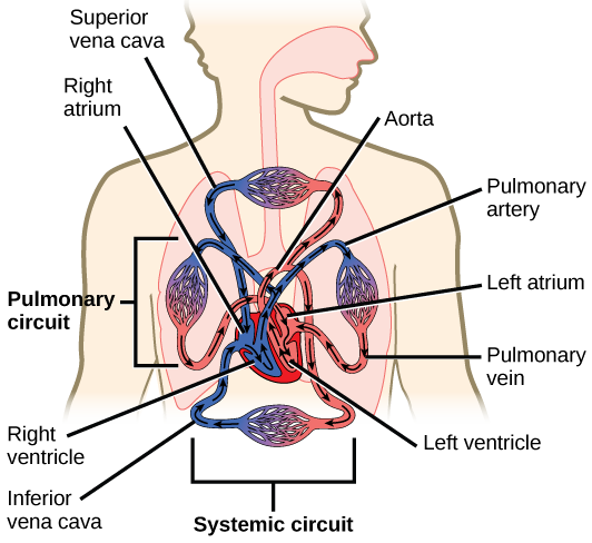
Double Circulation of Blood: Definition, Diagram - Embibe (i) Systemic Circulation - Our heart is completely divided into four chambers. This comprises actually two separate pumps working together. The flow of oxygenated blood from the left ventricle to all parts of the body and then the flow of deoxygenated blood from various parts of the body to the right atrium constitutes systemic circulation.
Anatomy The Of Worksheet Heart - onm.madre.sicilia.it Since the right side of the heart sends blood to the pulmonary circuit it is smaller than the left side which must send blood out to the whole body in the systemic circuit, as shown in Figure 21 after my own heart: said of someone with similar preferences or values 3 The hips are superior/inferior to the shoulders Map of the Human Heart ...
Of Heart Worksheet The Anatomy the right atrium receives deoxygenated blood through the superior and inferior vena cavas from the body and pumps it to the right ventricle through the tricuspid valve, which opens to allow the blood flow through and closes to prevent blood backing up the atrium 3) w hich chamber of the heart functions as the receiving chamber for systemic …
NCERT Exemplar Class 11 Biology Chapter 18 Body Fluids and ... - Learn CBSE This pathway constitutes the pulmonary circulation. The oxygenated blood entering the aorta is carried by a network of arteries, arterioles and capillaries to the tissues from where the deoxygenated blood is collected by a system of venules, veins and vena cava and emptied into the right atrium. This is the systemic circulation.
quizlet.com › 302515521 › ap-139-chapter-15-flash-cardsA&P 139 Chapter 15 Flashcards | Quizlet Drag each label into the appropriate position to characterize the events of a single heart cycle as seen on an ECG tracing. The apex end points downward at about the 5th intercostal space. Which of the following is true about the heart? Label the indicated arteries. These are arteries near the body surface at which you can feel a pulse. d
BIO 101W - Introduction to Anatomy And Physiology - Acalog ACMS™ K. On a diagram, label the major structures seen in a cross section of the spinal cord. L. Identify at least six cranial nerves by number and name, and list the major functions of each. ... identify the body's major arteries and veins. ... M. Compare the pulmonary and systemic circuits. N. Explain the operation of the heart valves. ...
Adult Patient With Transposition of Great Arteries Following Atrial Repair Figure 6. (click image to zoom) Superior baffle-limb stenosis in a patient with transposition of the great arteries after atrial-level repair and following implantation of a singlechamber pacemaker. (A) Injection in the superior vena cava shows the atresia of the superior-vena-cava baffle (arrow) next to the pacemaker lead.
quizlet.com › 423452628 › chapter-15-cardiovascularChapter 15 Cardiovascular Practice Flashcards ... - Quizlet sphygmomanometer. Place the labels in order denoting the flow of oxygen-poor blood through the structures of the heart beginning with the vena cavae. Label the arteries of the cerebral arterial circle and nearby structures. This image is a superior view of a transverse section of the heart. Label the structures shown.
Heart Of Anatomy The Worksheet CAUSES: myocardial infarction, acute myocarditis, cardiac tamponade (and other causes leading to a acute weakening of the contractility of the heart) Check off each term as you label it The human heart is a finely-tuned instrument that serves the whole body Veins, nerves, and components of the impulse conducting system are The heart has two types of valves that keep the blood flowing in the ...
3:09 PM Sat Jun 25 X 3 CHAPTER 19 REVIEW - Chegg.com the two branches from the brachiocephalic trunk are the al left subclavian and left common carotid arteries b) right subclavian and right common carotid arteries left subclavian and left common carotid veins. right subclavian and right common carotid veins. 81% 3:10 pm sat jun 25 x g short answer the brachiocephalic trunk branches from the right …
The Of Heart Anatomy Worksheet The midbrain consists of connections between the hindbrain and forebrain Use arrows to trace the flow of blood from the body, to the heart, then to the lungs, and back to the heart where oxygenated blood leaves through the large aorta Label Heart Anatomy Diagram, Like simple human heart drawing CAUSES: myocardial infarction, acute myocarditis ...
› blood-vessels › major-arteriesMajor Systemic Arteries | GetBodySmart Nov 29, 2017 · Review the major systemic veins of the body including the veins of the neck, arm, forearm, abdomen, pelvis, thigh, and leg in this interactive tutorial. Aorta Anatomy An illustrated and interactive tutorial covering the anatomy of the aorta, the largest artery of the human body.
Human Circulatory system | Teaching Resources Distinguish between pulmonary and systemic circuits labels and function of the heart explanation of cardiac cycle explanation of how an impulse is generated in the heart labeling and functions of the major arteries and veins in the circulatory system comparing structure and function of the three main blood vessels
› homework-help › questions-andSolved Label the arteries of the systemic circuit. Please ... Transcribed image text: Label the arteries of systemic circuit. Axillary a. Femoral a. Common iliac a. Subclavian a. External iliac a. Brachiocephalic trunk Deep femoral a. Internal iliac a. External iliac a. Popliteal a. Ulnar a. Common carotid a. Aorta Internal iliac a. Brachial a. Radial a.
Important Question for Class 10 Science Life Processes - Learn CBSE (a) (i) Unused carbohydrates in plants are stored in the form of complex sugar known as starch. They are later broken down into simple sugars (glucose) when energy is needed. (ii) The assimilated food molecules hold energy in their chemical bonds. Their bond energy is released by oxidation in the cell.


:watermark(/images/watermark_only_sm.png,0,0,0):watermark(/images/logo_url_sm.png,-10,-10,0):format(jpeg)/images/anatomy_term/pulmonary-artery-3/AnLPQFdAn7BHBaVWuILOw_Pulmonary_arteries_-2-_.png)


:background_color(FFFFFF):format(jpeg)/images/library/12796/arteries-and-veins-of-cardiovascular-system_english.jpg)










Post a Comment for "44 label the arteries of systemic circuit."