42 diagram of compound microscope with labelling
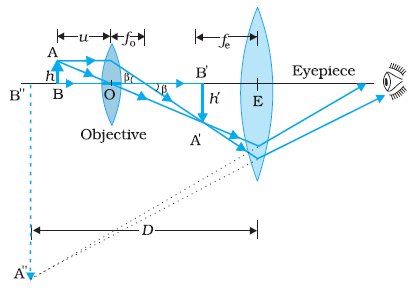
(b) Why both objective and eyepiece of a compound microscope must have ... (a) Draw the labelled ray diagram for the formation of image by a compound microscope. Derive an expression for its total magnification (or magnifying power), when the final image is formed at the near point. (b) Why both objective and eyepiece of a compound microscope must have short focal lengths? Compound Microscope: Definition, Diagram, Parts, Uses, Working ... - BYJUS The parts of a compound microscope can be classified into two: Non-optical parts Optical parts Non-optical parts Base The base is also known as the foot which is either U or horseshoe-shaped. It is a metallic structure that supports the entire microscope. Pillar The connection between the base and the arm are possible through the pillar. Arm
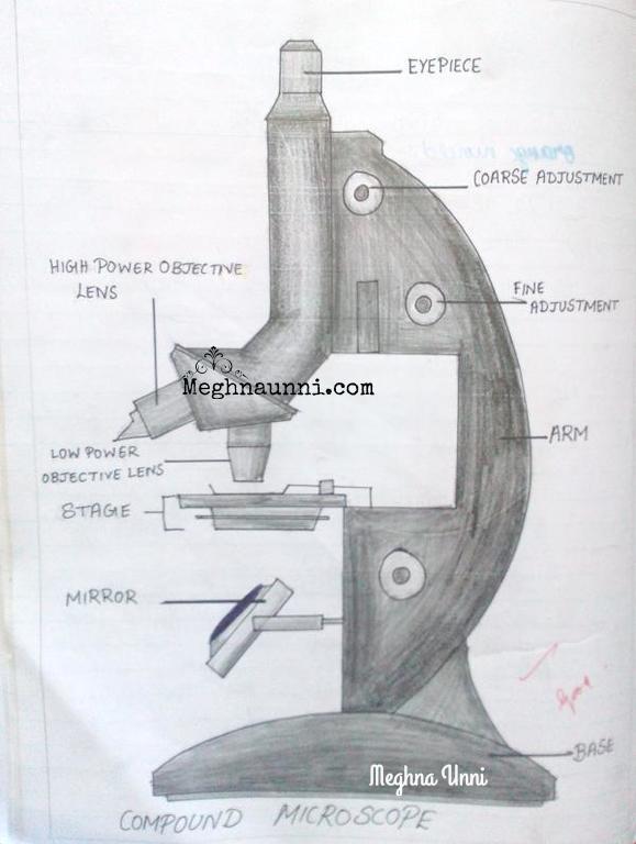
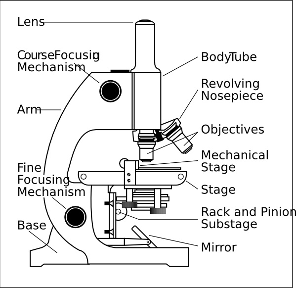
Labelled Diagram of Compound Microscope - Biology Discussion The below mentioned article provides a labelled diagram of compound microscope. Part # 1. The Stand: The stand is made up of a heavy foot which carries a curved inclinable limb or arm bearing the body tube. The foot is generally horse shoe-shaped structure (Fig. 2) which rests on table top or any other surface on which the microscope in kept.

Diagram of compound microscope with labelling
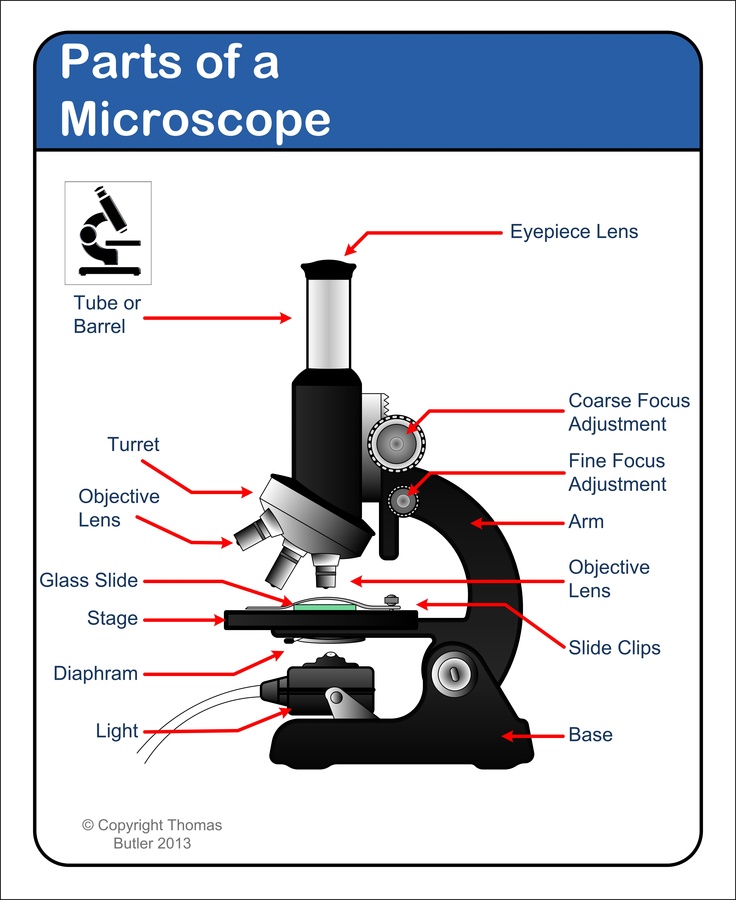
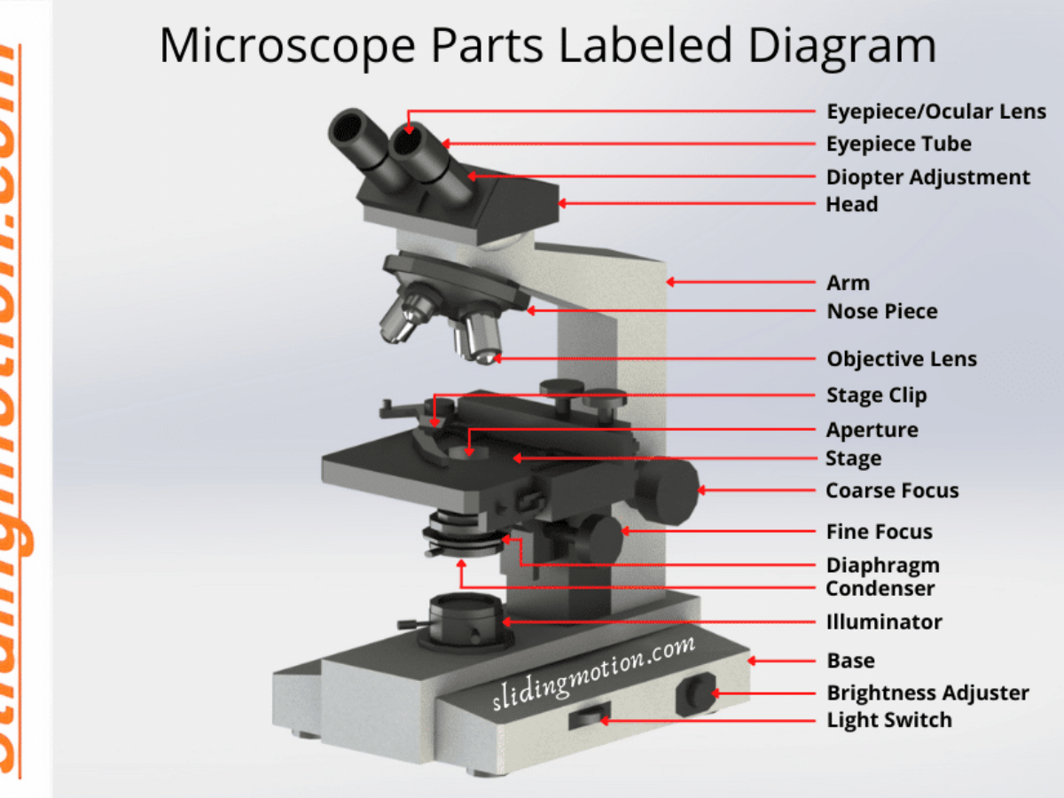
Microscope Parts, Function, & Labeled Diagram - slidingmotion Condenser. The condenser is to focus the light, which passes from the microscopic illuminator to the specimen. This condenser is located just below the diaphragm. This diaphragm is one of the important parts of the compound microscope which will help to get an accurate and sharp image. The condenser has a magnification power of 400X and above. Microscope Types (with labeled diagrams) and Functions Compound microscope labeled diagram Compound microscope functions: It finds great application in areas of pathology, pedology, forensics etc Its greater order of magnification allows for deeper study of microbial organisms to Detect the cause of diseases Study the mineral composition in soils Parts of the Microscope with Labeling (also Free Printouts) 5. Knobs (fine and coarse) By adjusting the knob, you can adjust the focus of the microscope. The majority of the microscope models today have the knobs mounted on the same part of the device. Image 5: The circled parts of the microscope are the fine and coarse adjustment knobs. Picture Source: bp.blogspot.com.
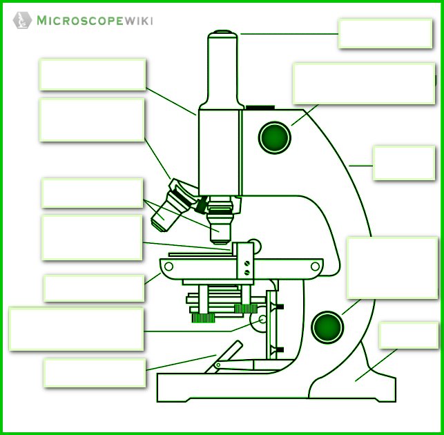
Diagram of compound microscope with labelling. Labeling the Parts of the Microscope Labeling the Parts of the Microscope This activity has been designed for use in homes and schools. Each microscope layout (both blank and the version with answers) are available as PDF downloads. You can view a more in-depth review of each part of the microscope here. Download the Label the Parts of the Microscope PDF printable version here. A Study of the Microscope and its Functions With a Labeled Diagram To better understand the structure and function of a microscope, we need to take a look at the labeled microscope diagrams of the compound and electron microscope. These diagrams clearly explain the functioning of the microscopes along with their respective parts. Man's curiosity has led to great inventions. The microscope is one of them. 16 Parts of a Compound Microscope: Diagrams and Video Once you have an understanding of the parts of the microscope it will be much easier to navigate around and begin observing your specimen, which is the fun part! The 16 core parts of a compound microscope are: Head (Body) Arm Base Eyepiece Eyepiece tube Objective lenses Revolving Nosepiece (Turret) Rack stop Coarse adjustment knobs Compound Microscope Parts – Labeled Diagram and their ... Labeled diagram of a compound microscope Major structural parts of a compound microscope There are three major structural parts of a compound microscope. The head includes the upper part of the microscope, which houses the most critical optical components, and the eyepiece tube of the microscope.
Draw a neat labelled diagram of a compound microscope and ... - Sarthaks Dividing and multiplying by I1 G1 on the right side, we get Magnifying power of the objective (m0) = I1G1/OJ = Height of the image due to the objective. Magnifying power of the eye piece (me) = IG/I1G1 = Height of the final image / Height of the object for the eyepiece. ∴ m = m0 × me ..... (1) Parts of a microscope with functions and labeled diagram Q. List down the 18 parts of a Microscope. 1. Ocular Lens (Eye Piece) 2. Diopter Adjustment 3. Head 4. Nose Piece 5. Objective Lens 6. Arm (Carrying Handle) 7. Mechanical Stage 8. Stage Clip 9. Aperture 10. Diaphragm 11. Condenser 12. Coarse Adjustment 13. Fine Adjustment 14. Illuminator (Light Source) 15. Stage Controls 16. Base 17. Diagram of a Compound Microscope - Biology Discussion The size of objects viewed under the compound microscope can be accurately determined using a micrometer. The latter consists of two scales, the eyepiece scale, (also called 'graticule' or 'ocular') and the stage micrometer scale. The eyepiece scale is calibrated with the help of stage micrometer and the former is then used for measurements. Microscope Labeling - The Biology Corner Students label the parts of the microscope in this photo of a basic laboratory light microscope. Can be used for practice or as a quiz. ... Microscope Labeling . Microscope Use: 15. When focusing a specimen, you should always start with the _____ objective. 16. When using the high power objective, only the _____ knob should be used. 17. The ...
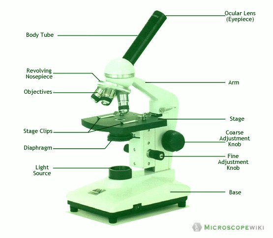
Compound Microscope Parts, Functions, and Labeled Diagram Nov 18, 2020 · Compound Microscope Definitions for Labels. Eyepiece (ocular lens) with or without Pointer: The part that is looked through at the top of the compound microscope. Eyepieces typically have a magnification between 5x & 30x. Monocular or Binocular Head: Structural support that holds & connects the eyepieces to the objective lenses. Microscope Labeling Diagram | Quizlet Animal Cell Labeling 1. 9 terms. PGFry210. Plant Cell Labeling 1. 10 terms. PGFry210. Animal Cell Labeling 2 ... Parts of a Microscope (Bio Quiz) 13 terms. angela2sweet. An Introduction to the Compound Microscope. 28 terms. MiracleFaith98. OTHER SETS BY THIS CREATOR. Unit 2 Lesson 7 - Biotechnology ... Diagrams. Flashcards. Mobile. Help. Sign ... Labeling the Parts of the Microscope | Microscope activity, Science ... Description Worksheet identifying the parts of the compound light microscope. Answer key: 1. Body tube 2. Revolving nosepiece 3. Low power objective 4. Medium power objective 5. High power objective 6. Stage clips 7. Diaphragm 8. Light source 9. Eyepiece 10. Arm 11. Stage 12. Coarse adjustment knob 13. Fine adjustment knob 14. Base S How to draw compound of Microscope easily - YouTube I will show you " How to draw compound of microscope easily - step by step "Please watch carefully and try this okay.Thanks for watching.....#microscopedrawi...
Compound Microscope Parts – Labeled Diagram and their ...
Parts of a Compound Microscope and Their Functions Compound microscope mechanical parts (Microscope Diagram: 2) include base or foot, pillar, arm, inclination joint, stage, clips, diaphragm, body tube, nose piece, coarse adjustment knob and fine adjustment knob. Base: It's the horseshoe-shaped base structure of microscope. All of the other components of the compound microscope are supported ...
Biology : Compound Microscope Diagram for Class 8 ...
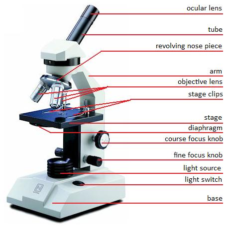
Compound Microscope- Definition, Labeled Diagram, Principle, Parts, Uses Parts of a Compound Microscope Eyepiece And Body Tube. The eyepiece is the lens through which the viewer looks to see the specimen. It usually contains a 10X or 15X power lens. The body tube connects the eyepiece to the objective lenses. Objectives and Stage Clips Objective Lenses are one of the most important parts of a Compound Microscope.

Labeling the Parts of the Microscope | Microscope World Resources
Compound microscope - their parts and function - Microscopy4kids 2. Eyepiece (10x) and Objective lenses (4x, 10x, 40x, 100x) are two major optical parts of a microscope. 3. Total magnification power is calculated by multiplying the magnification of the eyepiece and objective lens. 4. A proper immersion oil helps oil lens achieve an ideal magnification or resolution. 5.

How to draw Compound microscope | Microscope diagram easily Draw | SSC diagram
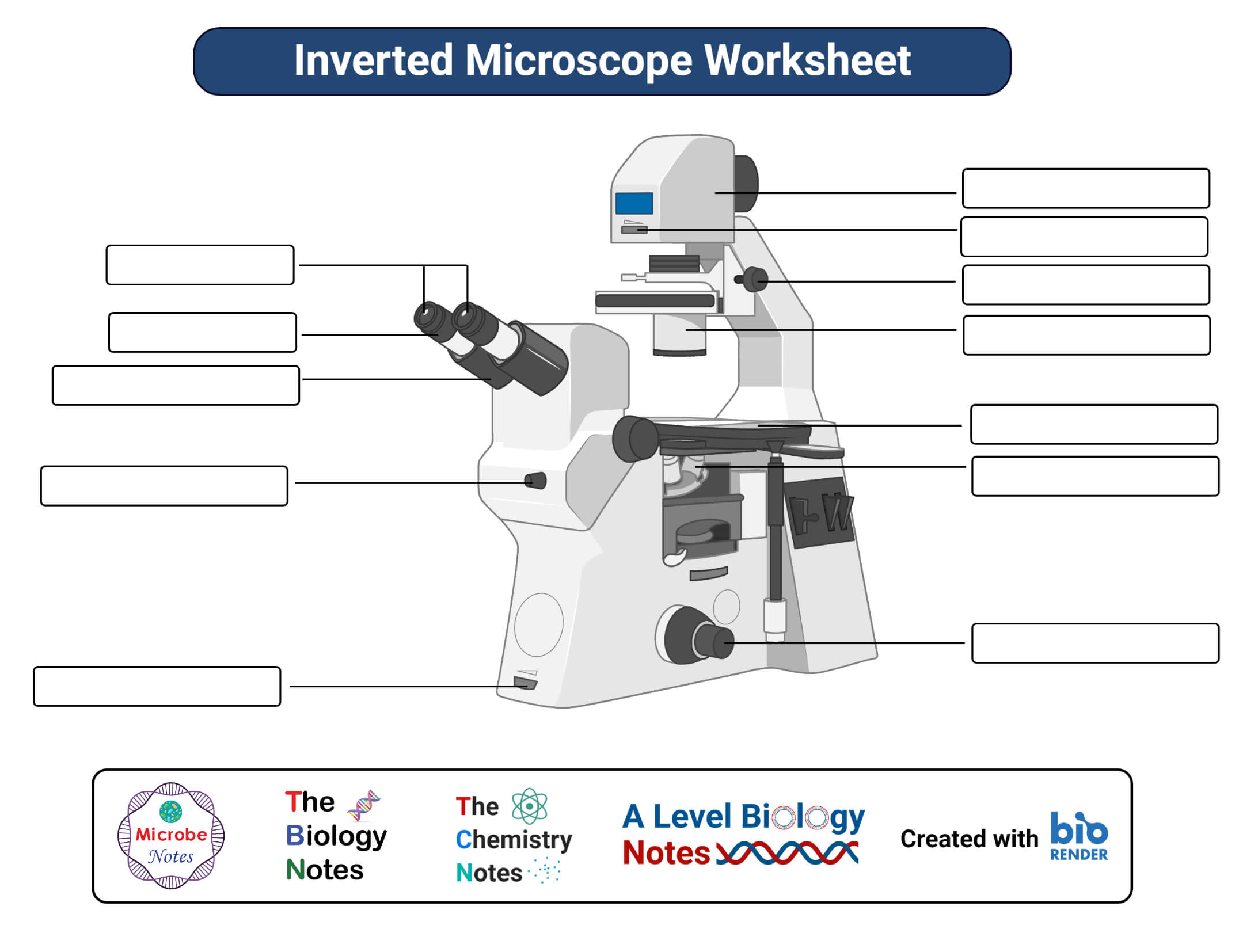
Compound Microscope - Types, Parts, Diagram, Functions and Uses It comes with a wide body and base. Its distinct parts include a condenser, illumination, focus lock, mechanical stage, and a revolving nosepiece which can hold up to five objectives. It usually has a binocular head, which makes long-term observation easy. Image 22: An example of a research compound microscope.

Parts of a Microscope - SmartSchool Systems
Label the microscope — Science Learning Hub All microscopes share features in common. In this interactive, you can label the different parts of a microscope. Use this with the Microscope parts activity to help students identify and label the main parts of a microscope and then describe their functions. Drag and drop the text labels onto the microscope diagram.

HOw to draw light or compound microscope step by step / Microscope diagram
Microscope Diagram and Functions | Science fair projects, Microscope ... A Study of the Microscope and its Functions With a Labeled Diagram. To better understand the structure and function of a microscope, we need to take a look at the labeled microscope diagrams of the compound and electron microscope. These diagrams clearly explain the functioning of the microscopes along with their respective parts.

Microscope With Labels Clip Art at Clker.com - vector clip ...
Parts of a Compound Microscope - Labeled (with diagrams) Parts of a Compound Microscope - Labeled (with diagrams) A compound microscope is known as a high-power microscope that enables you to achieve a high level of magnification. Smaller specimens can be thoroughly viewed using a compound microscope. Let us take a look at the different parts of a compound microscope and understand each key component.

Parts of a microscope with functions and labeled diagram
Compound Microscope Parts, Function, & Diagram | What is a Compound ... The base of the compound light microscope is the bottom portion of the compound microscope. It functions to support the entire compound microscope. The base can be set on a table or lab bench, and...
Label Microscope Parts - ClipArt Best
Compound Microscope – Diagram (Parts labelled), Principle and ... See: Labeled Diagram showing differences between compound and simple microscope parts Structural Components The three structural components include 1. Head This is the upper part of the microscope that houses the optical parts 2. Arm This part connects the head with the base and provides stability to the microscope.
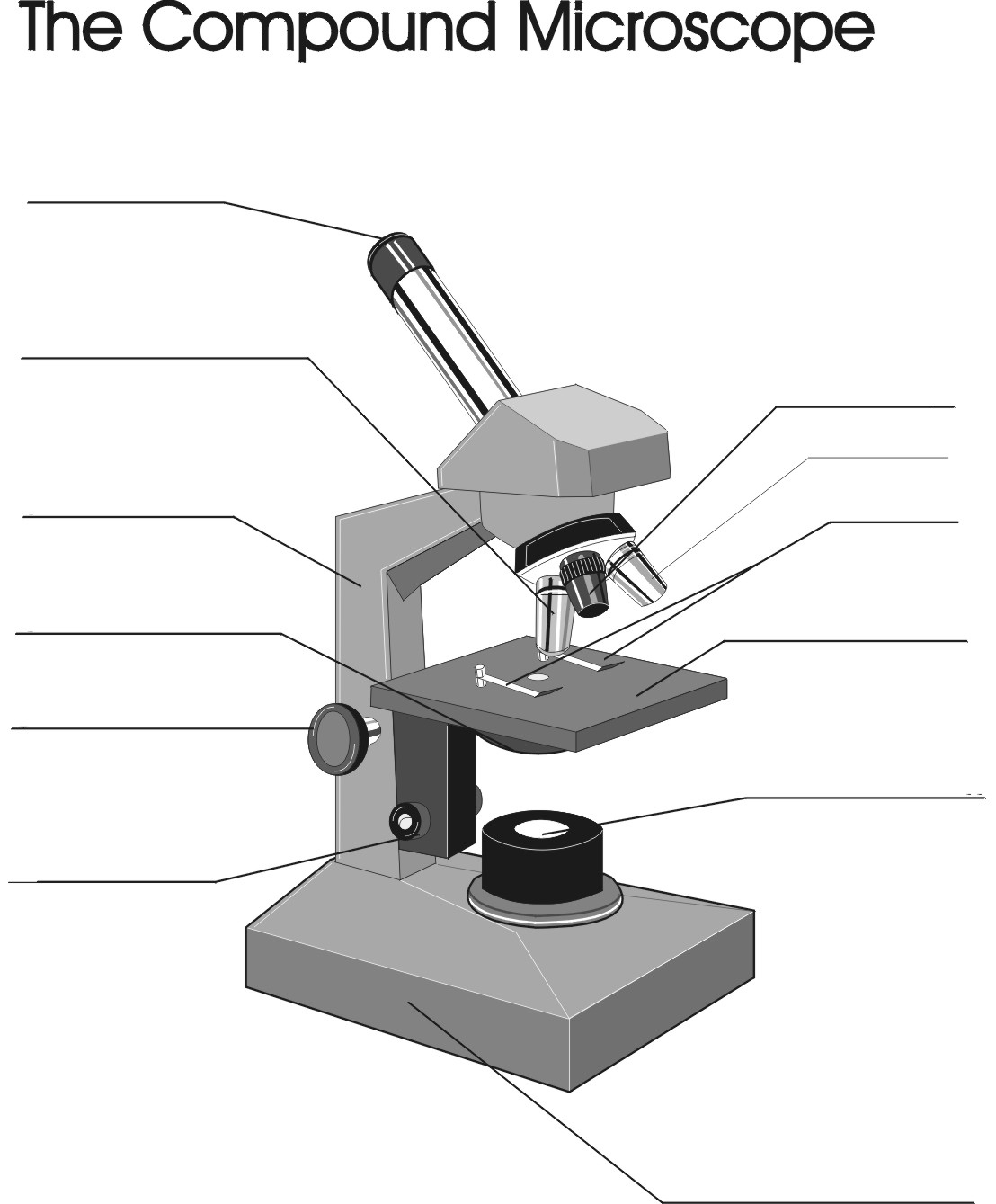
Remix of "The Compound Microscope"
Compound Microscope Labeled Diagram | Quizlet QUESTION. The total magnification of a specimen being viewed with a 10X ocular lens and a 40X objective lens is. 15 answers. QUESTION. a mosquito beats its wings up and down 600 times per second, which you hear as a very annoying 600 Hz sound. if the air outside is 20 C, how far would a sound wave travel between wing beats. 2 answers.
draw a labelled ray diagram showing imagr formation in ...
Microscope Parts and Functions With Labeled Diagram and ... Body tube (Head): The body tube connects the eyepiece to the objective lenses. Arm: The arm connects the body tube to the base of the microscope. Coarse adjustment: Brings the specimen into general focus. Fine adjustment: Fine tunes the focus and increases the detail of the specimen.
Microscope Types (with labeled diagrams) and Functions
Parts of the Microscope with Labeling (also Free Printouts) 5. Knobs (fine and coarse) By adjusting the knob, you can adjust the focus of the microscope. The majority of the microscope models today have the knobs mounted on the same part of the device. Image 5: The circled parts of the microscope are the fine and coarse adjustment knobs. Picture Source: bp.blogspot.com.

2.1 " Compound Microscope" | Download Scientific Diagram
Microscope Types (with labeled diagrams) and Functions Compound microscope labeled diagram Compound microscope functions: It finds great application in areas of pathology, pedology, forensics etc Its greater order of magnification allows for deeper study of microbial organisms to Detect the cause of diseases Study the mineral composition in soils

Biology - labeling a compound microscope Diagram | Quizlet
Microscope Parts, Function, & Labeled Diagram - slidingmotion Condenser. The condenser is to focus the light, which passes from the microscopic illuminator to the specimen. This condenser is located just below the diaphragm. This diaphragm is one of the important parts of the compound microscope which will help to get an accurate and sharp image. The condenser has a magnification power of 400X and above.

Parts of a Microscope Microscope Basics. Label the Compound ...
Free Microscope Drawing, Download Free Microscope Drawing png ...
Parts of a microscope with functions and labeled diagram

Parts of a Compound Microscope and Their Functions
Simple Microscope - Diagram (Parts labelled), Principle ...

Label the microscope — Science Learning Hub

Parts of Compound Microscope | Botany

Compound Microscope: Parts of Compound Microscope

Simple doodles, Microscope parts, Microscopic images

Microscope Parts and Functions

Microscope labeled diagram
Microscope With Labels clip art Vectors graphic art designs ...
biology labeled microscope diagram - Clip Art Library

Compound and Stereo Microscopes | Download Scientific Diagram
Parts of Microscope, Function, Names & Labeled Diagram ...
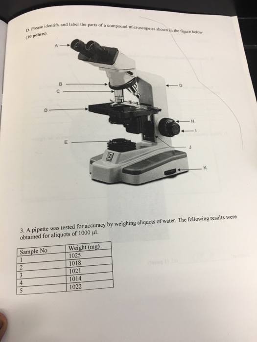
Solved Identify and label the parts of a compound microscope ...

Labelling a Microscope Diagram | Quizlet

Simple Microscope - Diagram (Parts labelled), Principle ...
Label a microscope - Teaching resources

Label the Microscope Diagram | Download Scientific Diagram

Compound Microscope Parts – Labeled Diagram and their ...

microscope diagram with name - EDUSIP

Label Microscope Diagram - EnchantedLearning.com

Compound Microscope Parts, Diagram Definition, Application ...

Draw a labelled diagram of a compound microscope.

This is a common compound microscope Label its parts class 11 ...
Microscope, Microscope Parts, Labeled Diagram, and Functions

Compound Microscope Parts, Functions, and Labeled Diagram ...
Post a Comment for "42 diagram of compound microscope with labelling"