42 label the internal anatomy of the heart.
Heart Anatomy Labeling Game - PurposeGames.com This is an online quiz called Heart Anatomy Labeling Game. There is a printable worksheet available for download here so you can take the quiz with pen and paper. Your Skills & Rank. Total Points. 0. Get started! Today's Rank--0. Today 's Points. One of us! Game Points. 19. You need to get 100% to score the 19 points available. Heart: Anatomy and Function - Cleveland Clinic Your heart walls are the muscles that contract (squeeze) and relax to send blood throughout your body. A layer of muscular tissue called the septum divides your heart walls into the left and right sides. Your heart walls have three layers: Endocardium: Inner layer. Myocardium: Muscular middle layer. Epicardium: Protective outer layer.
How to Draw the Internal Structure of the Heart (with Pictures) Finding a Diagram 1 To find a good diagram, go to Google Images, and type in "The Internal Structure of the Human Heart". Find an image that displays the entire heart, and click on it to enlarge it. 2 Find a piece of paper and something to draw with. Start with the pulmonary veins. They will be to the lower left of the Aorta. There are two of them.

Label the internal anatomy of the heart.
Heart Diagram with Labels and Detailed Explanation - BYJUS Diagram of Heart. The human heart is the most crucial organ of the human body. It pumps blood from the heart to different parts of the body and back to the heart. The most common heart attack symptoms or warning signs are chest pain, breathlessness, nausea, sweating etc. The diagram of heart is beneficial for Class 10 and 12 and is frequently ... › tag › anatomyAnatomy quizzes - PurposeGames Label the Heart by LMaggieO 1,454,796 plays 21p Image Quiz. ... Anatomy of the Human Heart - Internal Structures by orkide1 120,045 plays 24p Image Quiz. Correctly Label The Following Internal Anatomy Of The Heart The aorta, or aortic arch, is the outermost layer of the heart. The left ventricle is covered with the ventricular aorta, and the pulmonary veins are located inside the aorta. The two atria, the left and right aorta, and the right aortic arch are all external organs. These organs carry oxygen-rich blood to the body.
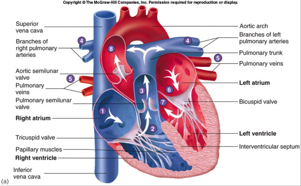
Label the internal anatomy of the heart.. Heart Anatomy: Labeled Diagram, Structures, Function, and Blood Flow We now have a 2x2 table in which we can label the boxes/chambers of the heart. Box 1: The first box is located in the right upper region. We know the atria are on top, and since box 1 is located on the right side, this is the right atrium. Box 2: The second box is also located on the right side, but now we are in the lower region. correctly label the following internal anatomy of the heart ... The epicardium is made of endocardium. Epicardium is the part of the heart that connects the myocardium and the endocardium. the epicardial wall of the left side of the heart is made of endocardium. The Epicardium is the internal structure of the heart. It is the first layer of the heart wall and is comprised of the endocardium. Internal Anatomy of a Whale with Label Stock Vector - Illustration of ... Illustration about Internal Anatomy of a Whale with label illustration. Illustration of alphabet, heart, living - 207562504 The Anatomy of the Heart, Its Structures, and Functions The heart is made up of four chambers: Atria: Upper two chambers of the heart. Ventricles: Lower two chambers of the heart. Heart Wall The heart wall consists of three layers: Epicardium: The outer layer of the wall of the heart. Myocardium: The muscular middle layer of the wall of the heart. Endocardium: The inner layer of the heart.
Human Heart (Anatomy): Diagram, Function, Chambers, Location in Body The heart is a muscular organ about the size of a fist, located just behind and slightly left of the breastbone. The heart pumps blood through the network of arteries and veins called the... Label the Heart - The Biology Corner Shows a picture of a heart with letters and blanks for practice with labeling the parts of the heart and tracing the flow of blood within the heart. › virtual › heartHeart Anatomy - Virtual Dissection - The Biology Corner Many anatomy units will include the dissection of the heart, and specimens can vary. Preserved sheep and cow hearts can be obtained from supply companies. Fresh hearts may also be available from local butchers or hunters. All mammalian hearts follow the same basic pattern: two atria, two ventricles, and four major vessels. Label the heart — Science Learning Hub In this interactive, you can label parts of the human heart. Drag and drop the text labels onto the boxes next to the diagram. Selecting or hovering over a box will highlight each area in the diagram. Right ventricle Right atrium Left atrium Pulmonary artery Left ventricle Pulmonary vein Semilunar valve Vena cava Aorta Download Exercise Tweet
The Heart - Science Quiz - GeoGuessr Aorta, Aortic valve, Left atrium, Left ventricle, Mitral valve, Pulmonary artery, Pulmonary valve, Pulmonary vein, Right atrium, Right ventricle, Septum, Superior vena cava, Tricuspid valve (13) Create custom quiz 0% | 0:05 | Click on: Aortic valve > Sound On Review PDF Internal Anatomy of Heart - Mrs. Hille's FunZone THE INTERNAL ANATOMY OF THE HEART Instructions: (1.) Read the statements. (2.) Use the statements to help you to label the diagram and answer the questions. RI Statements 1. Inside the heart are four spaces or chambers. Each chamber in the top half of the heart is called an "atrium"; the plural form is "atria." Arrows D and J point to the atria. Solved Correctly label the internal anatomy of the heart. | Chegg.com Question: Correctly label the internal anatomy of the heart. Tricuspid valve Left atrium Bicuspid valve Right atrium Aortic semilunar valve Pulmonary semilunar valve This problem has been solved! See the answer Show transcribed image text Expert Answer Solution : Heart is four chambered namely right and left a … View the full answer Heart Anatomy: Heart Dissection - University of Washington The major vessels of the heart are found at the base of the heart, along with the upper chambers, the right atrium (C) and left atrium (D). The atria are collapsed, but in a functioning heart, they would be stretched full of blood. The majority of the heart tissue consists of the ventricles. The left ventricle (F) is stiff and solid because it ...
Fetal Pig Anatomy Quiz
› internal-anatomy-of-an-insectColor Diagrams of Insect Organs and Internal Structures Jan 17, 2019 · A single blood vessel runs along the dorsal side of the insect, from the head to the abdomen. In the abdomen, the vessel divides into chambers and functions as the insect heart. Perforations in the heart wall, called ostia, allow hemolymph to enter the chambers from the body cavity.
Human Heart-Gross structure and Anatomy - Online Biology Notes
quizlet.com › 574029087 › ch-19-circulatory-systemCh. 19 Circulatory System- heart Flashcards | Quizlet Correctly label the following external anatomy of the posterior heart. Correctly label the external anatomy of the anterior heart. Place the labels in order denoting the flow of blood through the pulmonary circuit beginning with the right atrium and ending in the left atrioventricular valve.

Label the Heart Quiz
Internal anatomy of the heart (overview) | Internal anatomy of the ... Internally, the heart is divided into four chambers: right and left atria, and right and left ventricles. Functionally, the heart consists of two pumps, each consisting of an atrium and a ventricle separated by a valve: the right pump receives deoxygenated blood and pumps it into the lungs, and the left pump receives oxygenated blood from the lungs and sends it to the body.

SCIENCE
Human Heart - Diagram and Anatomy of the Heart - Innerbody The heart is a muscular organ about the size of a closed fist that functions as the body's circulatory pump. It takes in deoxygenated blood through the veins and delivers it to the lungs for oxygenation before pumping it into the various arteries (which provide oxygen and nutrients to body tissues by transporting the blood throughout the body).

Vertebrate Flashcards | Easy Notecards
Heart Labeling Quiz: How Much You Know About Heart Labeling? Here is a Heart labeling quiz for you. The human heart is a vital organ for every human. The more healthy your heart is, the longer the chances you have of surviving, so you better take care of it. Take the following quiz to know how much you know about your heart. Questions and Answers. 1.

fetal pig anatomy - YouTube
› worksheets › heart_internalLearn the Anatomy of the Heart - The Biology Corner Shows a picture of a heart with a description of how blood flows through the heart, focusing on the chambers, vessels, and valves. Students are asked to label the heart and trace the flow of blood. Questions at the end of the activity reinforce important concepts about the heart and circulatory system.

External Gross Anatomy of the Heart: Anterior View
19.1 Heart Anatomy - Anatomy & Physiology The position of the heart in the torso between the vertebrae and sternum (see Figure 19.1.1 for the position of the heart within the thorax) allows for individuals to apply an emergency technique known as cardiopulmonary resuscitation (CPR) if the heart of a patient should stop. By applying pressure with the flat portion of one hand on the sternum in the area between the line at T4 and T9 ...

Organs - Science Quiz
anatomyzone.com › abdomen-and-pelvisAbdomen and Pelvis - 3D Interactive Anatomy Tutorials 3D interactive models and tutorials on the anatomy of the abdomen and pelvis. Learn about the blood vessels, organs, nerves and peritoneal cavity.
Post a Comment for "42 label the internal anatomy of the heart."